Discover and publish cutting edge, open research.

Browse 69,679 multi-disciplinary research preprints

Featured documents

Michael Weekes

and 11 more

Nick K. Jones1,2*, Lucy Rivett1,2*, Chris Workman3, Mark Ferris3, Ashley Shaw1, Cambridge COVID-19 Collaboration1,4, Paul J. Lehner1,4, Rob Howes5, Giles Wright3, Nicholas J. Matheson1,4,6¶, Michael P. Weekes1,7¶1 Cambridge University NHS Hospitals Foundation Trust, Cambridge, UK2 Clinical Microbiology & Public Health Laboratory, Public Health England, Cambridge, UK3 Occupational Health and Wellbeing, Cambridge Biomedical Campus, Cambridge, UK4 Cambridge Institute of Therapeutic Immunology & Infectious Disease, University of Cambridge, Cambridge, UK5 Cambridge COVID-19 Testing Centre and AstraZeneca, Anne Mclaren Building, Cambridge, UK6 NHS Blood and Transplant, Cambridge, UK7 Cambridge Institute for Medical Research, University of Cambridge, Cambridge, UK*Joint first authorship¶Joint last authorshipCorrespondence: [email protected] UK has initiated mass COVID-19 immunisation, with healthcare workers (HCWs) given early priority because of the potential for workplace exposure and risk of onward transmission to patients. The UK’s Joint Committee on Vaccination and Immunisation has recommended maximising the number of people vaccinated with first doses at the expense of early booster vaccinations, based on single dose efficacy against symptomatic COVID-19 disease.1-3At the time of writing, three COVID-19 vaccines have been granted emergency use authorisation in the UK, including the BNT162b2 mRNA COVID-19 vaccine (Pfizer-BioNTech). A vital outstanding question is whether this vaccine prevents or promotes asymptomatic SARS-CoV-2 infection, rather than symptomatic COVID-19 disease, because sub-clinical infection following vaccination could continue to drive transmission. This is especially important because many UK HCWs have received this vaccine, and nosocomial COVID-19 infection has been a persistent problem.Through the implementation of a 24 h-turnaround PCR-based comprehensive HCW screening programme at Cambridge University Hospitals NHS Foundation Trust (CUHNFT), we previously demonstrated the frequent presence of pauci- and asymptomatic infection amongst HCWs during the UK’s first wave of the COVID-19 pandemic.4 Here, we evaluate the effect of first-dose BNT162b2 vaccination on test positivity rates and cycle threshold (Ct) values in the asymptomatic arm of our programme, which now offers weekly screening to all staff.Vaccination of HCWs at CUHNFT began on 8th December 2020, with mass vaccination from 8th January 2021. Here, we analyse data from the two weeks spanning 18thto 31st January 2021, during which: (a) the prevalence of COVID-19 amongst HCWs remained approximately constant; and (b) we screened comparable numbers of vaccinated and unvaccinated HCWs. Over this period, 4,408 (week 1) and 4,411 (week 2) PCR tests were performed from individuals reporting well to work. We stratified HCWs <12 days or > 12 days post-vaccination because this was the point at which protection against symptomatic infection began to appear in phase III clinical trial.226/3,252 (0·80%) tests from unvaccinated HCWs were positive (Ct<36), compared to 13/3,535 (0·37%) from HCWs <12 days post-vaccination and 4/1,989 (0·20%) tests from HCWs ≥12 days post-vaccination (p=0·023 and p=0·004, respectively; Fisher’s exact test, Figure). This suggests a four-fold decrease in the risk of asymptomatic SARS-CoV-2 infection amongst HCWs ≥12 days post-vaccination, compared to unvaccinated HCWs, with an intermediate effect amongst HCWs <12 days post-vaccination.A marked reduction in infections was also seen when analyses were repeated with: (a) inclusion of HCWs testing positive through both the symptomatic and asymptomatic arms of the programme (56/3,282 (1·71%) unvaccinated vs 8/1,997 (0·40%) ≥12 days post-vaccination, 4·3-fold reduction, p=0·00001); (b) inclusion of PCR tests which were positive at the limit of detection (Ct>36, 42/3,268 (1·29%) vs 15/2,000 (0·75%), 1·7-fold reduction, p=0·075); and (c) extension of the period of analysis to include six weeks from December 28th to February 7th 2021 (113/14,083 (0·80%) vs 5/4,872 (0·10%), 7·8-fold reduction, p=1x10⁻⁹). In addition, the median Ct value of positive tests showed a non-significant trend towards increase between unvaccinated HCWs and HCWs > 12 days post-vaccination (23·3 to 30·3, Figure), suggesting that samples from vaccinated individuals had lower viral loads.We therefore provide real-world evidence for a high level of protection against asymptomatic SARS-CoV-2 infection after a single dose of BNT162b2 vaccine, at a time of predominant transmission of the UK COVID-19 variant of concern 202012/01 (lineage B.1.1.7), and amongst a population with a relatively low frequency of prior infection (7.2% antibody positive).5This work was funded by a Wellcome Senior Clinical Research Fellowship to MPW (108070/Z/15/Z), a Wellcome Principal Research Fellowship to PJL (210688/Z/18/Z), and an MRC Clinician Scientist Fellowship (MR/P008801/1) and NHSBT workpackage (WPA15-02) to NJM. Funding was also received from Addenbrooke’s Charitable Trust and the Cambridge Biomedical Research Centre. We also acknowledge contributions from all staff at CUHNFT Occupational Health and Wellbeing and the Cambridge COVID-19 Testing Centre.

Guangming Wang

and 4 more

Tam Hunt

and 1 more

Tam Hunt [1], Jonathan SchoolerUniversity of California Santa Barbara Synchronization, harmonization, vibrations, or simply resonance in its most general sense seems to have an integral relationship with consciousness itself. One of the possible “neural correlates of consciousness” in mammalian brains is a combination of gamma, beta and theta synchrony. More broadly, we see similar kinds of resonance patterns in living and non-living structures of many types. What clues can resonance provide about the nature of consciousness more generally? This paper provides an overview of resonating structures in the fields of neuroscience, biology and physics and attempts to coalesce these data into a solution to what we see as the “easy part” of the Hard Problem, which is generally known as the “combination problem” or the “binding problem.” The combination problem asks: how do micro-conscious entities combine into a higher-level macro-consciousness? The proposed solution in the context of mammalian consciousness suggests that a shared resonance is what allows different parts of the brain to achieve a phase transition in the speed and bandwidth of information flows between the constituent parts. This phase transition allows for richer varieties of consciousness to arise, with the character and content of that consciousness in each moment determined by the particular set of constituent neurons. We also offer more general insights into the ontology of consciousness and suggest that consciousness manifests as a relatively smooth continuum of increasing richness in all physical processes, distinguishing our view from emergentist materialism. We refer to this approach as a (general) resonance theory of consciousness and offer some responses to Chalmers’ questions about the different kinds of “combination problem.”  At the heart of the universe is a steady, insistent beat: the sound of cycles in sync…. [T]hese feats of synchrony occur spontaneously, almost as if nature has an eerie yearning for order. Steven Strogatz, Sync: How Order Emerges From Chaos in the Universe, Nature and Daily Life (2003) If you want to find the secrets of the universe, think in terms of energy, frequency and vibration.Nikola Tesla (1942) I.               Introduction Is there an “easy part” and a “hard part” to the Hard Problem of consciousness? In this paper, we suggest that there is. The harder part is arriving at a philosophical position with respect to the relationship of matter and mind. This paper is about the “easy part” of the Hard Problem but we address the “hard part” briefly in this introduction.  We have both arrived, after much deliberation, at the position of panpsychism or panexperientialism (all matter has at least some associated mind/experience and vice versa). This is the view that all things and processes have both mental and physical aspects. Matter and mind are two sides of the same coin.  Panpsychism is one of many possible approaches that addresses the “hard part” of the Hard Problem. We adopt this position for all the reasons various authors have listed (Chalmers 1996, Griffin 1997, Hunt 2011, Goff 2017). This first step is particularly powerful if we adopt the Whiteheadian version of panpsychism (Whitehead 1929).  Reaching a position on this fundamental question of how mind relates to matter must be based on a “weight of plausibility” approach, rather than on definitive evidence, because establishing definitive evidence with respect to the presence of mind/experience is difficult. We must generally rely on examining various “behavioral correlates of consciousness” in judging whether entities other than ourselves are conscious – even with respect to other humans—since the only consciousness we can know with certainty is our own. Positing that matter and mind are two sides of the same coin explains the problem of consciousness insofar as it avoids the problems of emergence because under this approach consciousness doesn’t emerge. Consciousness is, rather, always present, at some level, even in the simplest of processes, but it “complexifies” as matter complexifies, and vice versa. Consciousness starts very simple and becomes more complex and rich under the right conditions, which in our proposed framework rely on resonance mechanisms. Matter and mind are two sides of the coin. Neither is primary; they are coequal.  We acknowledge the challenges of adopting this perspective, but encourage readers to consider the many compelling reasons to consider it that are reviewed elsewhere (Chalmers 1996, Griffin 1998, Hunt 2011, Goff 2017, Schooler, Schooler, & Hunt, 2011; Schooler, 2015).  Taking a position on the overarching ontology is the first step in addressing the Hard Problem. But this leads to the related questions: at what level of organization does consciousness reside in any particular process? Is a rock conscious? A chair? An ant? A bacterium? Or are only the smaller constituents, such as atoms or molecules, of these entities conscious? And if there is some degree of consciousness even in atoms and molecules, as panpsychism suggests (albeit of a very rudimentary nature, an important point to remember), how do these micro-conscious entities combine into the higher-level and obvious consciousness we witness in entities like humans and other mammals?  This set of questions is known as the “combination problem,” another now-classic problem in the philosophy of mind, and is what we describe here as the “easy part” of the Hard Problem. Our characterization of this part of the problem as “easy”[2] is, of course, more than a little tongue in cheek. The authors have discussed frequently with each other what part of the Hard Problem should be labeled the easier part and which the harder part. Regardless of the labels we choose, however, this paper focuses on our suggested solution to the combination problem.  Various solutions to the combination problem have been proposed but none have gained widespread acceptance. This paper further elaborates a proposed solution to the combination problem that we first described in Hunt 2011 and Schooler, Hunt, and Schooler 2011. The proposed solution rests on the idea of resonance, a shared vibratory frequency, which can also be called synchrony or field coherence. We will generally use resonance and “sync,” short for synchrony, interchangeably in this paper. We describe the approach as a general resonance theory of consciousness or just “general resonance theory” (GRT). GRT is a field theory of consciousness wherein the various specific fields associated with matter and energy are the seat of conscious awareness.  A summary of our approach appears in Appendix 1.  All things in our universe are constantly in motion, in process. Even objects that appear to be stationary are in fact vibrating, oscillating, resonating, at specific frequencies. So all things are actually processes. Resonance is a specific type of motion, characterized by synchronized oscillation between two states.  An interesting phenomenon occurs when different vibrating processes come into proximity: they will often start vibrating together at the same frequency. They “sync up,” sometimes in ways that can seem mysterious, and allow for richer and faster information and energy flows (Figure 1 offers a schematic). Examining this phenomenon leads to potentially deep insights about the nature of consciousness in both the human/mammalian context but also at a deeper ontological level.

Susanne Schilling*^

and 9 more

Jessica mead

and 6 more

The construct of wellbeing has been criticised as a neoliberal construction of western individualism that ignores wider systemic issues including increasing burden of chronic disease, widening inequality, concerns over environmental degradation and anthropogenic climate change. While these criticisms overlook recent developments, there remains a need for biopsychosocial models that extend theoretical grounding beyond individual wellbeing, incorporating overlapping contextual issues relating to community and environment. Our first GENIAL model \cite{Kemp_2017} provided a more expansive view of pathways to longevity in the context of individual health and wellbeing, emphasising bidirectional links to positive social ties and the impact of sociocultural factors. In this paper, we build on these ideas and propose GENIAL 2.0, focusing on intersecting individual-community-environmental contributions to health and wellbeing, and laying an evidence-based, theoretical framework on which future research and innovative therapeutic innovations could be based. We suggest that our transdisciplinary model of wellbeing - focusing on individual, community and environmental contributions to personal wellbeing - will help to move the research field forward. In reconceptualising wellbeing, GENIAL 2.0 bridges the gap between psychological science and population health health systems, and presents opportunities for enhancing the health and wellbeing of people living with chronic conditions. Implications for future generations including the very survival of our species are discussed.  

Mark Ferris

and 14 more

IntroductionConsistent with World Health Organization (WHO) advice [1], UK Infection Protection Control guidance recommends that healthcare workers (HCWs) caring for patients with coronavirus disease 2019 (COVID-19) should use fluid resistant surgical masks type IIR (FRSMs) as respiratory protective equipment (RPE), unless aerosol generating procedures (AGPs) are being undertaken or are likely, when a filtering face piece 3 (FFP3) respirator should be used [2]. In a recent update, an FFP3 respirator is recommended if “an unacceptable risk of transmission remains following rigorous application of the hierarchy of control” [3]. Conversely, guidance from the Centers for Disease Control and Prevention (CDC) recommends that HCWs caring for patients with COVID-19 should use an N95 or higher level respirator [4]. WHO guidance suggests that a respirator, such as FFP3, may be used for HCWs in the absence of AGPs if availability or cost is not an issue [1].A recent systematic review undertaken for PHE concluded that: “patients with SARS-CoV-2 infection who are breathing, talking or coughing generate both respiratory droplets and aerosols, but FRSM (and where required, eye protection) are considered to provide adequate staff protection” [5]. Nevertheless, FFP3 respirators are more effective in preventing aerosol transmission than FRSMs, and observational data suggests that they may improve protection for HCWs [6]. It has therefore been suggested that respirators should be considered as a means of affording the best available protection [7], and some organisations have decided to provide FFP3 (or equivalent) respirators to HCWs caring for COVID-19 patients, despite a lack of mandate from local or national guidelines [8].Data from the HCW testing programme at Cambridge University Hospitals NHS Foundation Trust (CUHNFT) during the first wave of the UK severe acute respiratory syndrome coronavirus 2 (SARS-CoV-2) pandemic indicated a higher incidence of infection amongst HCWs caring for patients with COVID-19, compared with those who did not [9]. Subsequent studies have confirmed this observation [10, 11]. This disparity persisted at CUHNFT in December 2020, despite control measures consistent with PHE guidance and audits indicating good compliance. The CUHNFT infection control committee therefore implemented a change of RPE for staff on “red” (COVID-19) wards from FRSMs to FFP3 respirators. In this study, we analyse the incidence of SARS-CoV-2 infection in HCWs before and after this transition.

How it works

Upload or create your research work
You can upload Word, PDF, LaTeX as well as data, code, Jupyter Notebooks, videos, and figures. Or start a document from scratch.
Disseminate your research rapidly
Post your work as a preprint. A Digital Object Identifier (DOI) makes your research citeable and discoverable immediately.
Get published in a refereed journal
Track the status of your paper as it goes through peer review. When published, it automatically links to the publisher version.
Learn More

Most recent documents

Abstract CSF-contacting neurons are involved in detecting changes in the cerebrospinal (CSF) circulation and recent studies report their role in nociception. Using neuronal tracers (Fluoro-Gold™, Cholera toxin subunit B, BDA), immunofluorescence (Anti-FG, Anti-OT-neurophysin, Anti-vasopressin, Anti-GABA, Anti-OTR) and electron microscopy, we describe oxytocinergic and vasopressinergic CSF-contacting neurons within the hypothalamic paraventricular nucleus (PVN) and their projections towards the rostral agranular insular cortex (RAIC). Our results show the presence of CSF-contacting neurons along the PVN that are labelled by oxytocin (OT) or/and vasopressin (AVP) and could secrete these peptides into CSF by dendritic projections. Besides, we report some oxytocinergic CSF-contacting neurons that send projections towards the RAIC. Inside the RAIC, our ultrastructural analysis shows that axons from PVN sustain synaptic connections with cortical GABAergic neurons that express oxytocin receptor (OTR) where we identify OT molecules as well. These findings support the possible role of CSF-contacting neurons in the neuronal modulation by releasing neuromodulators both at CSF and synaptic levels. Also, our results signal the extended means of oxytocinergic transmission, including its release inside RAIC promoting local GABAergic activity and its secretion towards CSF probably modulating many areas close to the ventricles, that can be involved in different conditions as nociception modulation.
Opiates are fast pain relievers that can cause respiratory arrest. I show new mechanisms how mu-opioids and high prenatal nicotine cause respiratory slowdown linked to slow wave sleep. Mu-opioids activate the medial habenula which activates the interpeduncular nucleus. The MHb-IPN system decreases respiration and alarm/arousal response to hypercapnia by projections to PAG, DRN, MRN and LPO→RMTg→vPAG. The same MHb-IPN circuit that causes respiratory slowdown, likely causes ventilatory deficits in mammalian neonates, known as sudden infant death syndrome (SIDS), linked to high fetal nicotine intake. Natural slowdown of respiration and heart rate is caused by slow wave sleep, when body is not moving. The MHb and rostromedial tegmental nucleus are known for high amount of mu-opioid receptors. Both were claimed to be activated by the MHb→IPN→MRN circuit that activates serotonin release, promotes slow SWS, rest, immune defense, recovery, sharp wave ripples, cortical spindles, replay of temporaly, spatialy and relationally bound memories, synaptogenesis and BDNF linked growth, but inhibits theta states, arousal, alert wakefulness, awareness and REM sleep linked circuits (Vadovičová, 2015). This updated circuit model explains role of the MHb→IPN→MRN→hippocampus + claustrum →cortical slow wave activity (SWA) in anesthesia, memory replay, loss of awareness, SWS and in theta states suppression. I propose new mechanisms for anesthetic ketamine and hallucinogens effect: activation of the IPN→MRN→claustrum→cortical SWA circuit by the 5-HT2a IPN and claustrum receptors. I show why are ketamine and hallucinogens anxiolytic and antidepressant, and how activation of 5-HT2a receptors in vACC/infralimbic cortex increases the safety, well-being signal, and cognitive flexibility.

ahmad matarneh

and 7 more

A rare case of Non-lupus full house nephropathy in a transplanted kidney, case report:Ahmad Matarneh(1), Omar Salameh(2), Sundus Sardar(1), Amanda Karasinski(1), Theja Channapragada(2), Muhammad Abdulbasit(1), Erik Washburn(3), , Nasrollah Ghahramani(1)Affiliations: 1. Department of nephrology, Penn state Milton S Hershey Medical Center, United States of America2. Department of Internal Medicine, Penn state Milton S Hershey Medical Center, United States of America3. Department of pathology, Penn state Milton S Hershey Medical Center, United States of AmericaCorresponding authorAhmad MatarnehAffiliation: Penn State health , Milton S.hershey medical centerContact: Email [email protected] # 717-708-6754Keywords:Autoimmune disorders, Systemic lupus erythematosus, Transplant rejection. Full house nephropathyAbstract:Non lupus full house nephropathy is a rare entity with an unclear incidence. It describes the kidney biopsy findings of positive deposits for IgG, IgA, IgM, C3, C1q on immunofluorescence in the absence of the classical diagnostic features of systemic lupus nephritis. This disease entity is becoming more recognised but further studies are still needed to evaluate the incidence, etiologies and management of this condition. Transplant glomerulopathy is a major cause for renal graft loss. It can present with a wide variety of manifestations; It can cause AKI, CKD or glomerular inflammations through an immune complex or autoimmune mediated damage.Introduction:Non lupus full house nephropathy is defined as having the classical findings of lupus nephritis which are positive deposits for IgG, IgA, IgM, C3, C1q on immunofluorescence but without the other extra-renal or seologic manifestations needed for SLE diagnosis i.e ACR criteria.(1) Systemic lupus erythematosus is a multi-system autoimmune disorder. It can involve several organs with variable presentations. The diagnosis of SLE is mainly based on establishing the presence of >= 4 points of the American College of Rheumatology criteria. (2) Lupus nephritis is the renal manifestation of SLE, it can have a wide range of manifestations which can include AKI, nephrotic or nephritic syndrome secondary to glomerulonephritis or it can ultimately lead to ESRD. Kidney biopsy remains the gold standard test to diagnose and to guide the treatment of lupus nephritis. Classical kidney involvement in SLE is based mainly on the pattern of histological involvement and it is divided into several classes according to the international society of nephrology. (3) Transplant rejection remains a huge challenge to both patients and treating physicians, it can result in variable degree of kidney dysfunction and ultimately it might lead to graft loss.(4) Several risk factors for transplant rejection have been described in literature and certainly, autoimmune diseases remain high on the list.(5) We hereby report a patient who presented with nephrotic syndrome and was found to have de-novo non lupus full house nephropathy in the transplanted kidney.Case presentation: Patient is a 57-year-old female with a past medical history of End-Stage Renal Disease that was managed with a living unrelated kidney transplant in 2016, Hypothyroidism, and Type 1 Diabetes Mellitus (T1DM) managed with an insulin pump. The patient underwent a living unrelated kidney transplant in 2016 with significant post-transplant complications with ureteral strictures. Initially, she was managed by nephrostomy followed by multiple indwelling stent exchanges. However, stents were repeatedly colonized with multiple bacterial and candida infections, requiring antibiotic and antifungal therapy. Due to recurrent acute infections mycophenolate mofetil was held and she had a stent removal in an attempt to decrease her risk of UTI’s.She presented to an outside hospital with a 1-week history of progressively worsening generalized headaches, nausea/vomiting, and elevated home blood pressures. Her blood pressure was noted to be 200/120. Initial labs were notable for hemoglobin of 8.5g/dl, blood urea nitrogen (BUN) of 82 mg/dL, and creatinine of 3.35mg/dL, with baseline creatinine 2.2-2.5mg/dL.Physical exam on presentation was significant for +1 lower extremity edema, with the remainder of the physical exam within normal limits, Patient was afebrile and BP was noted to be 171/81 mmHg.Investigations:Labs notable for potassium 6.1 mEq/L, chloride 116 mEq/L, bicarbonate 18 mmol/L, blood urea nitrogen (BUN) 82 mg/dL, creatinine 3.64 mg/dL, phosphorus 6.0 mg/dL, hemoglobin 8.7 g/dl, and platelets 148. VBG showed pH 7.240, PCO2 41, PO2 42, bicarbonate 17.6. Blood, urine, and fungal cultures were normal. A urine protein/creatinine (PCR) ratio was 12.74, consistent with nephrotic range proteinuria. (Table 1)Table 1. Laboratory investigations.Ultrasound retroperitoneal revealed hydronephrosis in the right lower quadrant, transplant kidney with urothelial and collecting system thickening, stable from the previous ultrasound one month prior. Given the patient’s history of prior urethral stricture, however, she had a right ureteral stent placed with the urology team. Following stent placement, however, the patient’s urine output was less than 200cc, and her creatinine rose to 3.9.An additional renal ultrasound with doppler was done to evaluate blood flow through the transplanted kidney, revealing increased renal artery resistive indices of 0.93 and associated high resistance arterial waveform, concerning for chronic rejection. While CMV/BK and donor-specific antibiotics were negative, Further immunological workup was done revealing positive Antinuclear Antibody (ANA) but negative anti-double stranded DNA (anti-dsDNA) and anti-smith antibody (Sm). C3 was borderline low and C4 was normal. Additional workup for membranoproliferative glomerulonephritis, including hepatitis B and C serologies, were negative. there were still concerns about rejection, and the patient underwent a renal biopsy with interventional radiology. The preliminary read from the biopsy was concerning T-cell kidney transplant rejection versus membranoproliferative glomerulonephritis versus lupus nephritis.The final read of the biopsy demonstrated membranoproliferative glomerulopathy, immune complex-mediated with rare cellular crescents, and foci of fibrinoid necrosis. The full-house immunofluorescence staining suggests lupus nephritis as a possible etiology. Mild tubulitis and interstitial mononuclear inflammation meet the criteria for borderline/suspicious for acute T-cell mediated rejection. (Figures 1-5)She was diagnosed as a case of chronic rejection with immune complex deposition disease. She was treated with 1 gram of intravenous methylprednisolone once and transitioned to 60 mg oral prednisone once daily throughout the remainder of the course and at discharge. Her BUN continued to rise in the setting of steroid use, as high as 178 mg/dL at discharge.. In addition, patient developed hypoalbuminemia with associated anasarca. Her anasarca was managed with a bumetanide drip that was transitioned to intermittent dosing of 2 mg bumetanide on discharge. The patient’s urine output increased to over 1L with diuretics, with creatinine levels down trending. On discharge, the patient’s creatinine was 2.98 mg/dL.Outcome:The patient was followed up in the transplant nephrology clinic two weeks after discharge. Creatinine improved to 2.63 mg/dL. Proteinuria decreased to 5.7 with a 50% reduction from hospital admission. Patient’s lower extremity swelling decreased significantly. She was maintained on tacrolimus 2 mg twice daily, MMF 500 mg twice daily. Given the biopsy findings and her improvement of her creatinine and proteinuria degree it was decided to continue treatment as an underlying chronic rejection.Discussion: Glomerulonephritis remains one of the leading causes of renal graft loss following kidney transplant.(6) The incidence is widely variable and differs between studies. Recurrent and de-novo glomerulonephritis following kidney transplant are not uncommon and have been described as a cause of renal graft loss and/or dysfunction. Recurrence of GN is typically more common than de-novo processes.(7) GN is one of the main causes of renal graft loss after kidney transplant, according to one of the studies it was noted that recurrence of GN was the third most common cause of renal graft loss. (8) De-novo disease is defined as any new disease process that occurs in the kidney graft unrelated to the primary kidney disease. De-novo disease can have a variable presentation as it can manifest as acute, subacute to chronic with manifestations usually arising from glomerulonephritis, tubulointerstitial nephritis or vascular disease. The incidence of de-novo GN is unclear and has been hypothesized to be occurring in 4-20% of transplant recipients.(9) Most common patterns of GN are FSGS, MGN, MPGN. (10) Transplant glomerulopathy remains one of the major debilitating conditions that transplant patients deal with. It can present with a chronic or acute rejection picture in the form of steady rise in creatinine and GN pattern of injury. (11) Systemic lupus nephritis is a chronic inflammatory autoimmune condition that can affect multiple organs with variable presentations. Diagnosis is usually based on the classical serologic and extra renal criteria. (12)Lupus nephritis is one of the serious manifestations of SLE, it is defined as the presence of significant proteinuria and classical findings on kidney biopsy. Classical kidney biopsy findings of lupus nephritis under light microscopy are I. minimal mesangial, II. Proliferative mesangial, III. Focal proliferative, IV. Diffuse proliferative, V. membranous and VI. Sclerosing. One of the relatively specific findings on direct immunofluorescence on kidney biopsy is finding deposits positive for IgG, IgA, IgM, C3 and C1q, a pattern that is usually referred to as the full house pattern. (13) This finding is relatively specific for lupus nephritis however, in order to establish the diagnosis of SLE, the ACR criteria must be met.(14) Non lupus nephritis describes the classical IF findings of LN but without the clinical or serological evidence of lupus. It is a newly developing entity and that means further studies are still needed to better understand the incidence and its exact pathophysiology and presentations. (15)Kidney transplant remains the gold standard treatment for ESRD. It has shown to improve both quality of life and survival in comparison to renal replacement therapies. (16) Transplant rejection occurs as a result of the body’s own immune system creating antibodies against the alloantigen from the transplanted kidney. (17) The response can be variable in intensity and onset, it ranges from hyperacute which can happen within minutes of transplant and is usually related to preformed antibodies or ABO mismatch. Acute rejection occurs any time following transplant and is subclassified depending on the underlying factor and immune mechanism to antibody mediated rejection (ABMR), which usually happens as a result of donor specific alloantibodies damaging the kidney causing peritubulitis/capillaritis. And acute T-cell mediated rejection (TCMR_ which is characterized by lymphocytic infiltrate into tubules, interstitium and arterial intima. Chronic rejection on the other hand usually occurs after 3 months and can be as a result of chronic T cell mediated or chronic antibody mediated. (18)Risk factors for transplant rejection include: HLA mismatch, Positive B cell crossmatchAdvanced age of the donor type of transplant and inadequate immunosuppression. (19) Patients with rejection present differently, any increase in creatinine more than 25% of the baseline or the presence / worsening of proteinuria should raise the suspicion for rejection. Kidney biopsy might be warranted if transplant rejection is suspected as it would guide the management and further prognosis. (20)There has been no cases in literature describing the presence of non-lupus-full house nephropathy in the setting of post kidney transplant, we hypothesize that it might be related to circulating immune complex mediated damage to the transplant kidney possibly in the setting of chronic rejection. The point of interest in this condition and in our case in particular is that the patient was treated mainly for the possible underlying rejection with resuming mycophenolate and optimizing tacrolimus with institution steroids which lead to improvement in her kidney parameters and more than 50% drop in the degree of proteinuria. This further suggests that the possible underlying mechanism is related to the rejection process causing the non lupus pattern and nephrotic syndrome.Conclusion: Non lupus full house nephropathy is rare and is poorly understood. It has been described in association to several conditions however, There has been no data describing the associated post kidney transplant and rejection. Non lupus full house nephropathy can occur in the setting of kidney transplant and it might be related to transplant rejection. We wanted to raise awareness about this condition and possible association in transplant recipients as it carried out a worse prognosis and can lead to loss of graft function, also, to describe the methods in the diagnosis and a proposed treatment regimen.References:• Silva MD, Oliveira PV, Vale PH, Cunha RD, Lages JS, Brito DJ, Salgado Filho N, Guedes FL, Silva GE, Santos RF. Non-lupus full-house nephropathy: a case series. Brazilian Journal of Nephrology. 2020 Nov 11;43:586-90. • Mok CC, Lau CS. Pathogenesis of systemic lupus erythematosus. Journal of clinical pathology. 2003 Jul;56(7):481. • Ayoub I, Cassol C, Almaani S, Rovin B, Parikh SV. The kidney biopsy in systemic lupus erythematosus: a view of the past and a vision of the future. Advances in chronic kidney disease. 2019 Sep 1;26(5):360-8. • Vaillant AA, Mohseni M. Chronic Transplantation Rejection. InStatPearls [Internet] 2023 Jan 1. StatPearls Publishing. • Neuberger J. Incidence, timing, and risk factors for acute and chronic rejection. Liver transplantation and surgery: official publication of the American Association for the Study of Liver Diseases and the International Liver Transplantation Society. 1999 Jul 1;5(4 Suppl 1):S30-6. •Allen PJ, Chadban SJ, Craig JC, Lim WH, Allen RD, Clayton PA, Teixeira-Pinto A, Wong G. Recurrent glomerulonephritis after kidney transplantation: risk factors and allograft outcomes. Kidney international. 2017 Aug 1;92(2):461-9.• • Jafari R, Mehrazma M, Vahedi M, Ossareh S. Prevalence and Prognosis of Post-transplant Glomerulonephritis in Kidney Transplant Biopsies, A Single-Center Report. Iranian Journal of Kidney Diseases. 2023 Mar 1;17(2):92. • Briganti EM, Russ GR, McNeil JJ, Atkins RC, Chadban SJ. Risk of renal allograft loss from recurrent glomerulonephritis. New England Journal of Medicine. 2002 Jul 11;347(2):103-9. • Abbas F, El Kossi M, Jin JK, Sharma A, Halawa A. De novo glomerular diseases after renal transplantation: How is it different from recurrent glomerular diseases?. World Journal of Transplantation. 2017 Dec 12;7(6):285. • Hariharan S, Adams MB, Brennan DC, Davis CL, First MR, Johnson CP, Ouseph R, Peddi VR, Pelz CJ, Roza AM, Vincenti F. RECURRENT AND DE NOVO GLOMERULAR DISEASE AFTER RENAL TRANSPLANTATION: A Report from Renal Allograft Disease Registry (RADR): 1, 2. Transplantation. 1999 Sep 15;68(5):635-41. • Filippone EJ, McCue PA, Farber JL. Transplant glomerulopathy. Modern Pathology. 2018 Feb 1;31(2):235-52. • Gill JM, Quisel AM, Rocca PV, Walters DT. Diagnosis of systemic lupus erythematosus. American family physician. 2003 Dec 1;68(11):2179-87. • Musa R, Brent LH, Qurie A. Lupus nephritis. • Anders HJ, Saxena R, Zhao MH, Parodis I, Salmon JE, Mohan C. Lupus nephritis. Nature reviews Disease primers. 2020 Jan 23;6(1):7. • Wani AS, Zahir Z, Gupta A, Agrawal V. Clinicopathological pattern of non-lupus full house nephropathy. Indian Journal of Nephrology. 2020 Sep;30(5):301. • Braun MM, Khayat M. Kidney Disease: End-Stage Renal Disease. FP essentials. 2021 Oct 1;509:26-32. • Halloran PF, Einecke G, Sikosana ML, Madill-Thomsen K. The biology and molecular basis of organ transplant rejection. InPharmacology of Immunosuppression 2022 Jan 29 (pp. 1-26). Cham: Springer International Publishing. • Wood KJ, Goto R. Mechanisms of rejection: current perspectives. Transplantation. 2012 Jan 15;93(1):1-0. • Oweira H, Ramouz A, Ghamarnejad O, Khajeh E, Ali-Hasan-Al-Saegh S, Nikbakhsh R, Reißfelder C, Rahbari N, Mehrabi A, Sadeghi M. Risk factors of rejection in renal transplant recipients: a narrative review. Journal of Clinical Medicine. 2022 Mar 3;11(5):1392. • Williams WW, Taheri D, Tolkoff-Rubin N, Colvin RB. Clinical role of the renal transplant biopsy. Nature Reviews Nephrology. 2012 Feb;8(2):110-21.Acknowledgments: We thank the nephrology department at penn state health for giving us the opportunity and support to conduct this workAuthor contribution: Ahmad Matarneh: Manuscript writing, literature review, Clinical careOmar salameh Manuscript writing, literature review, Clinical careSundus sardar Manuscript writing, literature review, Clinical careTheja Channapragada Manuscript writing, literature review, Clinical careMohammad Abdulbasit Manuscript writing, literature review, Clinical careAmanda Karasinsk iManuscript writing, literature review, Clinical careErik Washburn HistopathologyNasrollah ghahramani: Clinical care, literature review, manuscript write up, MentorConsent: Verbal informed consent was obtained from the patient to publish this report in accordance with the journal’s patient consent policyConflict of interest The authors associated with the this case report have no actual or possible conflict of interest to declareDate availability statement: The data that support the findings of this study are available on request from the corresponding authorFunding: The funding process is solely done from the writing authors
The Deep Reef Refugia Hypothesis suggests that disturbances decrease with increasing depth, thus, reefs at the mesophotic zone potentially serve as refuges for communities found in shallower areas. This study aimed to evaluate fish diversity at shallow and mesophotic reefs at Parque Nacional Espíritu Santo (PNZMAES) and compare them to determine if deep reefs indeed serve as refuges for shallow communities. During 2021-2022 we conducted five-minute video-transects using remotely operated vehicles to document species richness and abundance at shallow and mesophotic habitats. For the recorded species, six biological traits were compiled (length, mobility, position, gregariousness, diet, and activity period) to estimate four functional indices (number of entities, richness, originality, and divergence) as well as Hill’s numbers for taxonomic and functional indices. Furthermore, monthly water turbidity (Kd490) satellite data products were transformed into a light attenuation coefficient (KdPAR) to locate the mesophotic zone (area between 10 and 0.1% of light penetration). At PNZMAES, the mesophotic zone was identified to extend to 21 m under optimal conditions (high stratification and low primary production), which is shallower compared to what is typically observed in oligotrophic regions. The PERMANOVA revealed significant variations in reef fish composition across spatial (site and zone) and temporal (season and year) dimensions. Additionally, univariate tests of functional richness, originality, and taxonomic Hill’s numbers exhibited significantly higher values in the shallow zone. However, functional divergence and functional Hill’s numbers indicated similarities in fish assemblages. Despite differences in fish taxonomic diversity among zones that could be related to less habitat complexity, environmental variation, and resource availability at deep strata, mesophotic reef fish assemblages presented similar functions. This maintenance of functions at mesophotic reefs suggests connectivity between zones and their potential role as a partial refugia in the face of current and near-future disturbances that could affect shallow zones.

Christian Mazimpaka

and 10 more

Introduction: Disrespect and abuse during childbirth represents a pervasive issue worldwide. In Rwanda, however, research in this area remains scarce. This study evaluated the factors linked to disrespect and abuse during labor and delivery in two Rwandan district hospitals. Methods: Employing a mixed method, cross-sectional design, we studied 280 women giving birth at Kabutare and Kibagabaga hospitals. Analysis was conducted with SPSS version 22, using the Chi-square test to identify factors correlated with childbirth-related disrespect and abuse. Results: Our findings revealed that 27% of women reported verbal abuse and 27.5% reported abandonment by healthcare providers, with 12% experiencing physical abuse during childbirth. Factors increasing the likelihood of experiencing disrespect and abuse included residing in rural areas [AOR=4.06, 95% CI (1.12, 14.7); p=0.03], having only primary education [AOR=2.9, 95% CI: 1.10-8.9, p=0.04], and night-time delivery [AOR=2.23, 95% CI (1.34, 3.03); p=0.03]. However, having a cesarean delivery [AOR=0.23, 95% CI: 0.09-0.59, p=0.002] or having attended antenatal care [AOR=0.28, 95% CI: 0.13-0.81, p=0.029] significantly reduced the risk of such experiences. Conclusion: The study uncovers a high incidence of disrespect and abuse during labor and delivery in Rwanda. The identified risk factors, namely rural residence, lower education, and night-time delivery, highlight specific areas requiring targeted intervention. The results advocate for effective measures to ensure dignified and equitable maternity care for all women.

Browse more recent preprints

Powerful features of Authorea

Under Review
Communities
Collections
Learn More
Journals connected to Under Review
Ecology and Evolution
Allergy
Clinical Case Reports
Land Degradation & Development
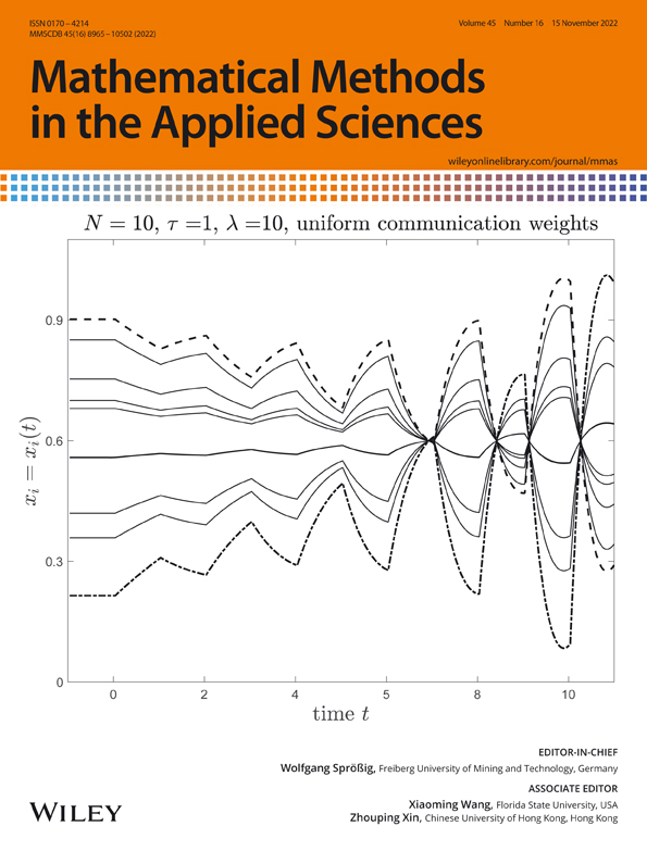
Mathematical Methods in the Applied Sciences
Biotechnology Journal
Plant, Cell & Environment
International Journal of Quantum Chemistry
PROTEINS: Structure, Function, and Bioinformatics
All IET journals
All AGU journals
All Wiley journals
READ ABOUT UNDER REVIEW
Featured Collection
READ ABOUT COLLECTIONS
Featured communities
Explore More Communities

Other benefits of Authorea

Multidisciplinary

A repository for any field of research, from Anthropology to Zoology

Comments

Discuss your preprints with your collaborators and the scientific community

Interactive Figures

Not just PDFs. You can publish d3.js and Plot.ly graphs, data, code, Jupyter notebooks

Documents recently accepted in scholarly journals

Karma Norbu

and 3 more

Introduction: Scrub typhus is a neglected life threatening acute febrile illness caused by bacteria Orientia tsutsugamushi and it is a vector-borne zoonotic disease. In 2009, scrub typhus outbreak at Gedu has awakened Bhutan on the awareness and testing of the disease.Information and data of the study highlights the need for in depth surveillance, awareness among prescribers and initiate preventive measures in the country. Methods: We used retrospective descriptive study through review of laboratory registers across three health centres in Zhemgang district, south central Bhutan. The laboratories registers have been transcribed into CSV file using Microsoft excel. Variables of interest were collected from the registers and then analysed using open statistical software R, (R Core Team (2020). R: A language and environment for statistical computing. R Foundation for Statistical Computing, Vienna, Austria.) And use of mStats package, (MyoMinnOo (2020). mStats: Epidemiological DataAnalysis. R package version 3.4.0.) Results: Of the total 922 tests prescribed for suspected scrub typhus in the three health centers in Zhemgang, only 8.2 % (n=76) were tested positive. Of these, Panbang Hospital had highest reported positive for scrub typhus with 56.6 %( n=43) followed by Yebilaptsa Hospital 35.5 %( n=27) and Zhemgang Hospital with 7.9 %( n=6). The female gender is comparably more affected as opposed to male with 57.9% (n=44) of the positive cases being female. The prevalence of scrub typhus seems to be affected by the seasonal variation as the months of Spring, Summer and Autumn together accounts for 98.7%(n=75) of total positive cases. The year 2019 noted significant scrub typhus cases accounting to 89.5 %(n=68) of the total positive cases over the two years. Conclusions:The overall tests tested positive of the scrub typhus infection within two years was 8.2%.

Browse more published preprints

Featured templates
Featured and interactive
Journals with direct submission
Explore All Templates