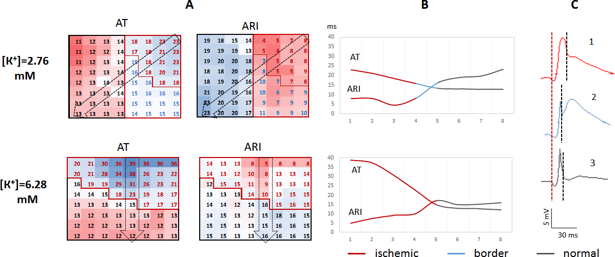
In the border zone of ischemic myocardium, extracellular potassium concentration ([K+]) gradually decreases from the ischemic to normal area. Since blood [K+] is equilibrated with the normal tissue [K+], it might affect the [K+] and modify electrophysiological properties in the border zone. The study aimed at evaluation of distribution of depolarization and repolarization characteristics across the ischemic-normal border under blood [K+] variation. 64-lead epicardial mapping was performed in 26 anesthetized rats with [K+] ranged from 2.3 to 6.4 mM in an in vivo model of acute ischemia/reperfusion. The animals with [K+]<4.7 mM (low-normal potassium) had a typical ischemic zone with ST-segment elevation and activation delay, a border zone with ST-segment elevation and no activation delay, and a normal zone without electrophysiological abnormalities. The animals with [K+] >4.7 mM (normal-high potassium) had only the typical ischemic and normal zones and no transitional area. Activation-repolarization intervals and local conduction velocities were inversely associated with [K+] in linear regression analysis with the adjustment for a zone of myocardium. The reperfusion extrasystolic burden (ESB) was greater in the low-normal as compared to normal-high potassium animals. Ventricular tachycardia/fibrillation incidence did not differ between the groups. In patch-clamp experiments, hypoxia shortened action potential duration at 5.4 mM but not at 1.3 mM of [K+]. The IK(ATP) current was lower at 1.3 mM than at 5.4 mM of [K+]. The formation of the border zone was associated with attenuation of IK(ATP) response to hypoxia in low-normal [K+] and increased ESB at reperfusion.