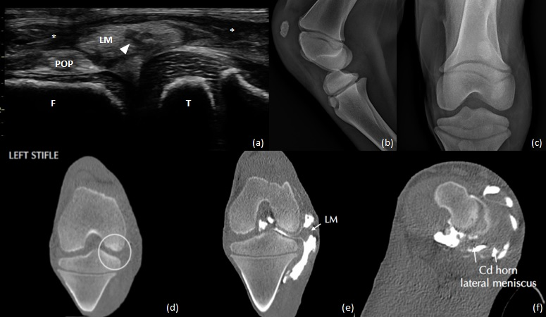
Objective: To report clinical characteristics, surgical management, and medium-term outcomes of 3 Arabian neonatal foals with meniscal disruption associated with septic arthritis of the lateral femorotibial joint. Methods: Three neonatal Arabian foals with septic arthritis of the lateral femorotibial joint (LFTJ), were diagnosed with lateral meniscal (LM) tears, based on persistent lameness despite improving synovial parameters, ultrasound (US) findings (protrusion of meniscal tissue beyond the level of the condyles, with hypoechoic regions), contrast Computed Tomography findings, and confirmed on arthroscopy. Treatment included arthroscopic debridement and lavage of the joint with debridement of the meniscal tear. Postoperative care included systemic and intra-articular antimicrobials, based on culture and sensitivity results. Two of the foals received intra-articular injections of autologous mesenchymal stem cells. Results: Grade III meniscal tears were observed in the LFTJ of the affected joints of all foals, involving the meniscal body (n=3) and caudal horn (n=1). Purulent material within the torn tissue, was debrided with a synovial resector. Foal 1 was lame-free as a yearling. Foal 2 was lame at walk at 7.5 months and euthanatized due to poor prognosis. Foal 3 showed mild lameness at trot in a straight line at 6 months. Disruption of the LM continued to be visible on US in both foals at these time-points. Conclusion: Meniscal disruption and infection should be considered a differential in neonatal foals with persistent femorotibial septic arthritis. In such cases, the LM could be the primary nidus of infection.