Nirupama Trehanpati

and 8 more

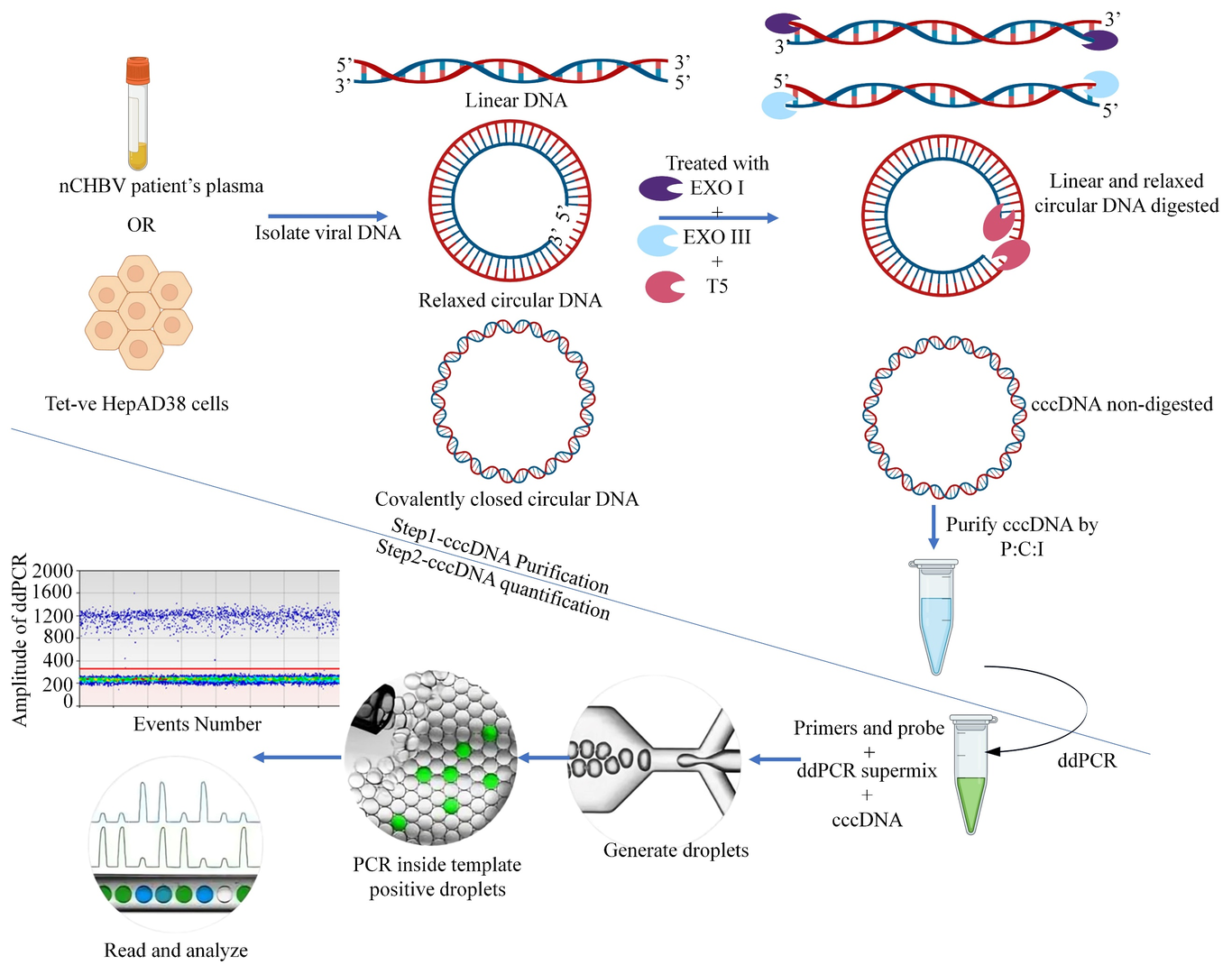
Covalently closed circular DNA (cccDNA) is a stable, episomal form of HBV DNA. cccDNA is a true marker for the intrahepatic events in controlled CHB infection. Quantifying cccDNA is critical for monitoring disease progression and efficacy of anti-viral therapies. To standardize the method, total HBV DNA was isolated from HepAD38 cells and digested with three exonuclease enzymes to remove linear and relaxed circular HBV DNA. Purified cccDNA quantification used ddPCR with specific primers. Treatment-naive chronic hepatitis B virus patients (nCHBV, n=36) with detectable HBV DNA and HBsAg, were grouped by HBsAg levels: Group I (HBsAg lo < 2000 IU/ml, n=11) and Group II (HBsAg hi > 2000 IU/ml, n=25). cccDNA, HBV DNA and HBsAg, were quantified in plasma and compared between groups. Correlation with clinical/histopathological features was done. Non-digested 3.6 ^10 6 tet -ve HepAD38 cells showed 316 copies/µl of total viral DNA. After digesting the linear, integrated, and relaxed circular DNA with triple enzymes, 15 copies/µl of cccDNA were detected. Similarly, after DNA digestion, HBsAg lo patients showed a median of 8.5 copies/µl (IQR 2.75-9.75 copies/µl), and HBsAg hi gave a median of 11 copies/µl (IQR 4-16 copies/µl) but with no significant difference between groups (p=0.093). Further, HBsAg lo patients with low cccDNA copy numbers showed significantly higher fibrosis grades than HBsAg hi (p=0.036). We conclude that employing a combined approach utilizing three exonucleases, cccDNA-specific primers, and ddPCR enables the detection of cccDNA copies even in patients exhibiting low levels of HBsAg and HBV DNA. This integrated method offers additional validation as a surrogate diagnostic tool.

Baibaswata Nayak

and 10 more

Background: Hepatitis B virus (HBV) infection is one of the major causes of chronic liver disease which progresses from hepatitis to liver fibrosis, cirrhosis and hepatocellular carcinoma (HCC). Early detection and laboratory based screening test of HCC is still a major challenge. HBV induces hepatocarcinogenesis through viral genome integration, chromosomal aberrations and modulation of host signaling pathways. Molecular alterations of cancer hallmark genes occur during the process of hepatocarcinogenesis. These signatures may release into the circulation through . circulating tumor DNA (ctDNA). Detection of these mutations in ctDNA may serve as liquid biopsy marker for screening, early detection and prognosis of HCC for which this study was undertaken. Methods: Consecutive patients of CHB-HCC (n=80), chronic hepatitis B (n=35) and healthy (n=15) controls were included for blood sample collection. The ctDNA was isolated from serum. Amplification and sequencing TP53 exon 7 and β-catenin exon 3 was carried out for predominant mutations in the ctDNA of HCC patients. Highly sensitive dual-probe based droplet digital PCR (ddPCR) assays were performed for TP53 (p.R249M & p.R249S) and β-catenin (p.S45P) driver mutations in healthy, CHB-noncirrhotic, cirrhotic and HCC patients. Results: Both TP53 gene exon 7 and CTNNB1 gene exon 3 region was amplified and sequenced in 32 HCC patient whereas sensitive ddPCR assay TP53 (p.R249M & p.R249S) and β-catenin (p.S45P) mutation for all 130 subjects. In sanger sequencing TP53 c.746 G˃T, p.R249M mutation was predominant. In ddPCR assay, 58.75% of HCC patients (n=47) ctDNA had at least one driver mutation in the ctDNA. Combined TP53 and CTNNB1 mutation was observed in 12.5% of HCC patients. Increased mutation frequency was observed in CHB-cirrhotic. CHB-HCC than CHB-noncirrhotic and healthy subjects. Percentage mutant fraction was highest in CHB-HCC than only CHB patients. Significant association TP53. R249M with smoking was observed in CHB-HCC patients. Poor survival was observed in HCC patients with combined TP53 and CTNNB1 gene mutation. Conclusion: Driver mutation screening for TP53 and CTNNB1 gene can be done in ctDNA for early diagnosis and prognosis.