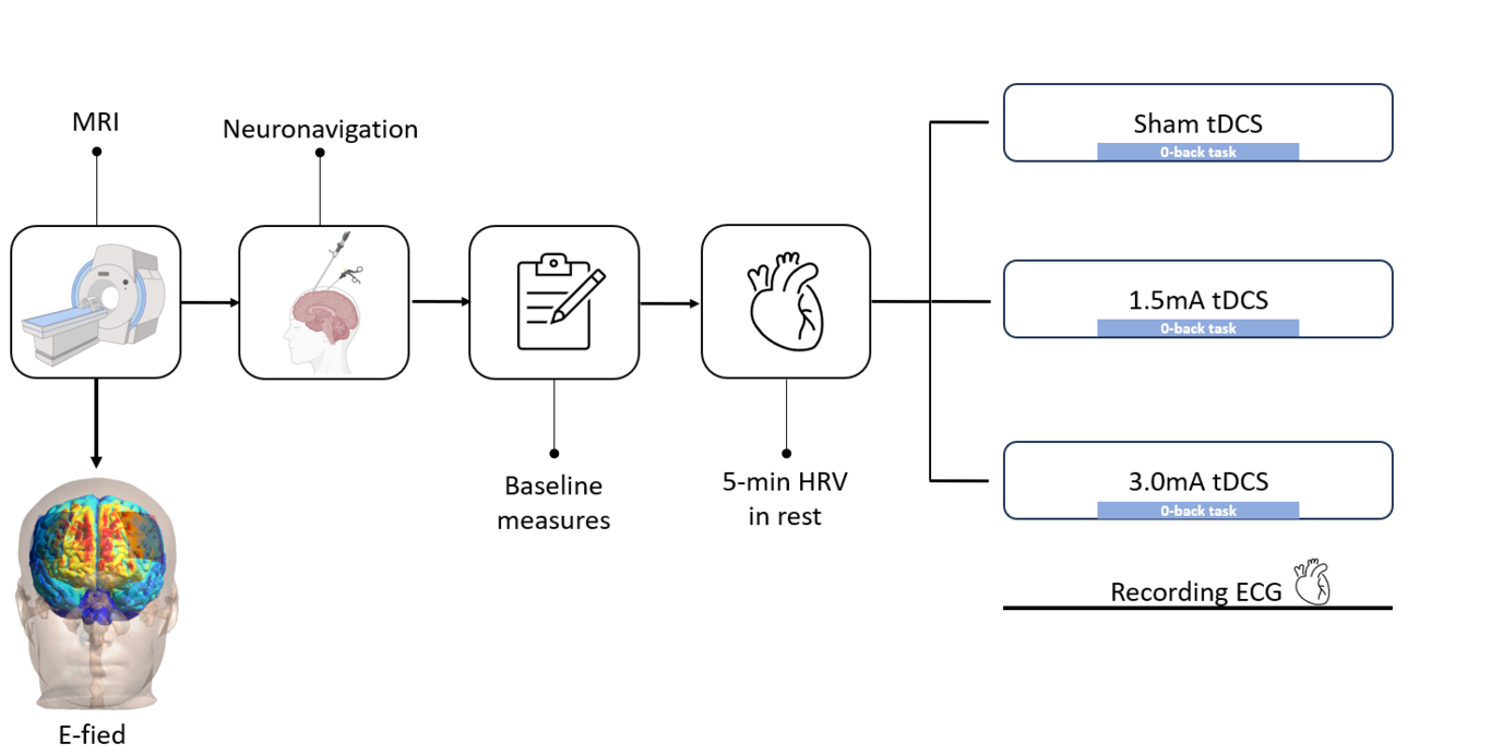
Transcranial direct current stimulation (tDCS) of the prefrontal cortex (PFC) modulates the autonomic nervous system by activating deeper brain areas via top-down pathway. However, effects on the nervous system are heterogeneous and may depend on the amount of current that penetrates the brain due to individual brain anatomical differences. Therefore, investigated the variable effects of tDCS on heart rate variability (HRV), a measure of the functional state of the autonomic nervous system. Using three prefrontal tDCS protocols (1.5mA, 3mA and sham), we associated the simulated individual electric field (E-field) magnitude in brain regions of interest with the HRV effects. This was a randomized, double-blinded, sham-controlled and within-subject trial, in which participants received tDCS sessions separated by two weeks. The brain regions of interest were the dorsolateral PFC (DLPFC), anterior cingulate cortex, insula and amygdala. Overall, 37 participants (mean age = 24.3 years, standard deviation = 4.8) were investigated, corresponding to a total of 111 tDCS sessions. The findings suggested that HRV, measured by Root Mean Squared of Successive Differences (RMSSD) and high-frequency HRV (HF-HRV), were significantly increased by the 3.0mA tDCS when compared to sham and 1.5mA. No difference was found between sham and 1.5mA. E-field analysis showed that all brain regions of interest were associated with the HRV outcomes. However, this significance was associated with the protocol intensity, rather than inter-individual anatomical variability. To conclude, our results suggest a dose-dependent effect of tDCS for HRV. Therefore, further research is warranted to investigate the optimal current dose to HRV.