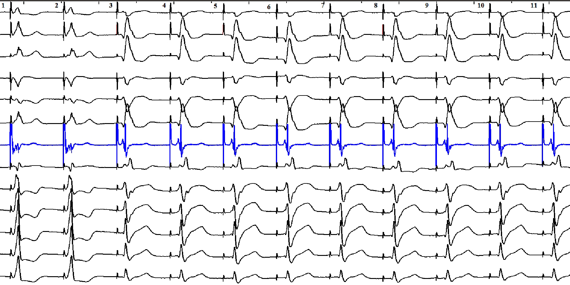
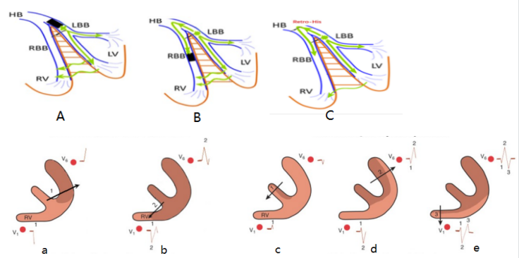
This study analyzed the characteristics of electrocardiograms recorded before electrode screwing and at the site of lead fixation during LBBP. The study aims to establish an electrocardiographic vector-guided method for electrode implantation during LBBP. Methods: 140 patients who underwent successful LBBP and had ECGs recorded before electrode screwing (pre-screwing) and at the site of lead fixation (post-implantation) were included in the study. Patients whose post-implantation ECGs revealed rSR’ or M morphologies in lead V1 were enrolled in the typical right bundle branch (RBBB) group, and those whose ECGs revealed qR, QR, Qr or QS morphologies in V1 were enrolled in the atypical RBBB group. Electrocardiographic characteristics of pre-screwing and post-implantation ECGs were thoroughly analyzed. Results: For the electrocardiograms of typical RBBB group, pre-screwing R-wave amplitudes in lead I, aVL, V4, and V5 were 0.6635 (0.433,0.866), 0.69 (0.487,0.866), 0.6635 (0.514,1.489), and 1.056 (0.677,1.840)mv, respectively; post-implantation QRS transition zone was 1 (0.5,3), and Stim-LVAT was 68 (61,74)ms. For the atypical RBBB group, pre-screwing R-wave amplitudes in lead I, aVL, V4 and V5 were 0.527 (0.379,0.704), 0.5285 (0.2985,0.785), 0.5545 (0.2165,1.11), and 0.8525 (0.4737,1.42), respectively; post-implantation QRS transition zone was 2 (1,3), and Stim-LVAT was 76 (65,81)ms. The between-group difference were statistically significant for all the parameters. Conclusion: In pre-screwing electrocardiograms, R-wave amplitude ≥0.35mV in aVL is a highly sensitive indicator for predicting typical RBBB morphologies in left bundle branch pacing, and R-wave amplitude≥0.7mV in lead I is an indicator with high specificity.