Larry Lands

and 3 more

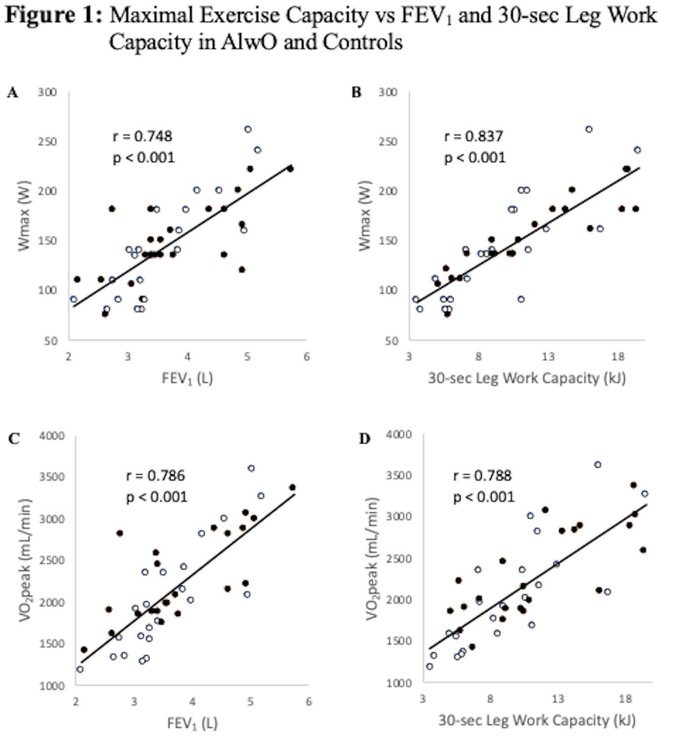
Background Adolescents living with obesity (AlwO) can have limited exercise capacity. Exercise capacity can be predicted by a 2-factor model comprising lung function and leg muscle function, but no study has looked at cycling leg muscle function and its contribution to cycling exercise capacity in AlwO. Methods 22 nonobese adolescents and 22 AlwO (BMI>95 percentile) were studied. Anthropometry, body composition (DEXA), spirometry, 30-sec isokinetic work capacity, and maximal exercise (cycle ergometry) were measured. Results AlwO had greater lean leg mass (LLM) (14.8±4.1 vs 21.0±4.3 kg, Con vs AlwO p<0.001). Lung function did not differ, although FEV 1 trended higher in AlwO (101.0±13.1 vs 107.9±12.7 percent predicted, p=0.08). Leg 30-second work output did not differ in absolute terms or per allometrically scaled LLM. Peak oxygen consumption did not differ between the groups in absolute terms or as percent predicted values (78.5±15.4 vs 82.1±16.5 percent predicted), but was lower in AlwO when expressed per scaled kg of LLM. Peak oxygen consumption related to both lung function and 30-second work capacity, with no observed group effect. 30-second leg work capacity related to the scaled muscle mass, with a small group effect. There was some correlation between leg work capacity and time spent in moderate to vigorous physical activity in AlwO (r s=0.39, p=0.07). Conclusion AlwO have larger LLM and preserved exercise capacity, when expressed as percentage of predicted, but not per allometrically scaled LLM. Increasing time spent in moderate to vigorous activity may benefit AlwO.

Noah Marzook

and 6 more

Background: Lung ultrasound (LUS) has been shown to be an effective tool to rapidly diagnose certain causes of pediatric respiratory distress in the emergency department. However, very little is known about LUS findings in pediatric asthma outside of acute exacerbations. Objectives: The primary objective of this study was to characterize LUS findings in a cohort of pediatric patients with a definitive diagnosis of asthma, outside of an asthma exacerbation. Methods: Eligible patients, aged 6 to 17 years old and diagnosed with asthma, underwent LUS during an outpatient visit. LUS was conducted using a six-zone scanning protocol. A positive LUS was defined by one or more of the following: ≥3 B-lines per intercostal space, pulmonary consolidation and/or pleural anomaly. Images were interpreted by an expert sonographer blinded to patient clinical characteristics. Results: 52 patients were included. 10/52 patients had a positive LUS (19.2%, 95CI 8.3-30.1%). Of those with positive LUS findings, 8 had B lines, 7 had consolidations <1cm, 1 had a pleural line abnormality and 1 had a consolidation >1cm. Positive findings were seen in the right anterior and lateral zones in 60% of participants and were limited to 1-2 intercostal spaces within one lung zone in 100% of participants. Conclusion: To our knowledge, this is the first report of LUS findings in outpatient pediatric asthma. Positive LUS findings in asthmatic children can be seen outside of acute exacerbations. Such findings need to be taken into consideration when using LUS for the acute evaluation of a pediatric patient with asthma.