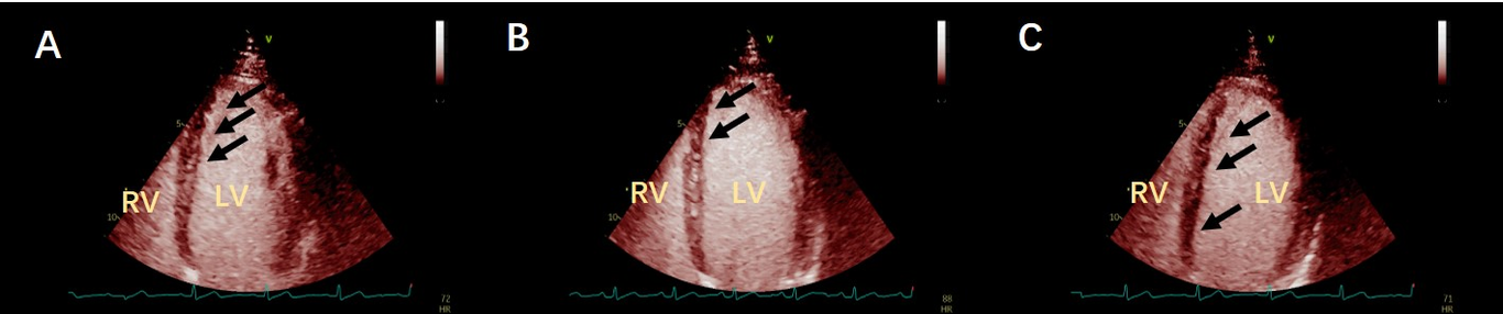
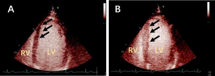
We report a case of coronary microvascular spasm assessed by ATP stress MCE (myocardial contrast electrocardiography). The patient had chest pain, but the coronary angiography was normal. There was apical ventricular septal perfusion delay before ATP stress, and the perfusion was significantly improved at peak stress, which was similar to the radionuclide myocardial perfusion characteristics of coronary microvascular spasm, In the recovery period, the flow spectrum resistance of the distal coronary artery of the left anterior descending artery increased compared with that before stress, which further confirmed that local coronary microvascular spasm was induced after vasodilation.