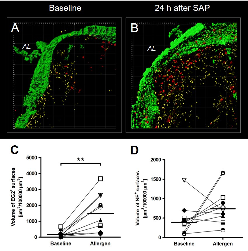
Allergic inflammation after allergen challenge – insights from the tissueTo the Editor,Limited data exist on the infiltration of eosinophils in direct response to allergen exposure in asthmatic patients. The experimental procedure of segmental allergen provocation (SAP) in mild asthmatic subjects is an extremely valuable study tool to investigate mechanisms of bronchial asthma in patients in general and in particular for the role of eosinophils. In this procedure, BAL and bronchial mucosa can be analysed simultaneously after the induction of allergic inflammation. Older studies yielded data on different time points after the challenge with contradictory results. Eosinophils were studied in mucosa and airway lumen of mild asthmatics undergoing segmental allergen provocation1,2. Here, eosinophils and their release products were investigated in thick sections of the bronchial biopsies. Subject data, methods and detailed results are given in the supplement. All subjects showed a clear eosinophilic response in the airway lumen and mucosa 24 hours after the challenge (Figure 1, S1). The data on neutrophils were inconclusive (Figure 1, S2). There was an increase of eosinophils in the mucosa and in the BAL. In the BAL the concentrations of IL‑5 and ECP were elevated (supplement). In the mucosa ECP stained areas were found elevated as well as signs of eosinophil activation and degranulation (Figure 2). ECP staining was associated with cellular structures, small granules or dispersed over a large area beneath the epithelium. The degree of released mediators into the tissue is an important parameter investigating new drugs for asthma. In the presented study not cell associated, free ECP positive granules were seen at baseline but only to a small extent. However, after allergen challenge the ECP volume was significantly increased and only few distinct cells could be detected being positive for ECP. These results suggest that in human tissue eosinophils degranulate to a significant amount after a single allergen challenge and therefore release all their toxic content into the surrounding tissue. This is in contrast to animal models of asthma where eosinophilic degranulation after allergen challenge does not occur extensively 3,4. In the present study, subjects with a strong IL-5 reaction in BAL showed a strong eosinophilic response in the lumen. However, BAL IL-5 levels did not correlate with eosinophil numbers in the mucosa or volume of ECP positive surfaces in the mucosa (Figure S3). It is a long known fact that IL-5 levels in the BAL correlate highly with absolute numbers of eosinophils in the same compartment 5. In the present study the allergic reaction is comparable to those of other studies. The missing correlation between IL-5 in the BAL and numbers of tissue eosinophils showed that the relationship between both compartments is not as simple as assumed. Here, the data for the volume of ECP positive surfaces may give an important hint. Interestingly, the volume of ECP positive surfaces was inversely correlated with TNF-α and IL-8. TNF-α enhances migration of eosinophils from mucosa to lumen shown in an in vitro cell culture model 6. Therefore, one possible explanation is the more TNF-α is found in the BAL the less activated eosinophils are found in the mucosa. In conclusion, the findings in subjects with mild asthma are in alignment with other published results and suggest that 1) human tissue eosinophils release their granules in non-provoked state, and 2) toxic content of these cells is significantly released into the surrounding tissue after a single allergen challenge whereas the distribution and the degree of activation and degranulation of eosinophils differs widely between subjects. Many eosinophils in the airways indicate many eosinophils in the mucosa. Three-dimensional analysis in thick tissue sections using confocal microscopy is a valuable tool in the investigation of bronchial biopsies from patients suffering from bronchial asthma.(Abbreviations: BAL=bronchoalveolar lavage, ECP=eosinophilic cation protein, IL=interleukin, MBP=major basic protein, NE=neutrophilic elastase, SAP=segmental allergen provocation, TNF=tumor necrosis factor)References1. Erpenbeck VJ, Hagenberg A, Dulkys Y, et al. Natural porcine surfactant augments airway inflammation after allergen challenge in patients with asthma. Am J Respir Crit Care Med.2004;169(5):578-586.2. Schaumann F, Muller M, Braun A, et al. Endotoxin augments myeloid dendritic cell influx into the airways in patients with allergic asthma.Am J Respir Crit Care Med. 2008;177(12):1307-1313.3. Denzler KL, Borchers MT, Crosby JR, et al. Extensive eosinophil degranulation and peroxidase-mediated oxidation of airway proteins do not occur in a mouse ovalbumin-challenge model of pulmonary inflammation. J Immunol. 2001;167(3):1672-1682.4. Malm-Erjefält M, Persson CG, Erjefält JS. Degranulation status of airway tissue eosinophils in mouse models of allergic airway inflammation. Am J Respir Cell Mol Biol. 2001;24(3):352-359.5. Sur S, Kita H, Gleich GJ, Chenier TC, Hunt LW. Eosinophil recruitment is associated with IL-5, but not with RANTES, twenty-four hours after allergen challenge. J Allergy Clin Immunol. 1996;97(6):1272-1278.6. Kikuchi I, Kikuchi S, Kobayashi T, et al. Eosinophil trans-basement membrane migration induced by interleukin-8 and neutrophils. Am J Respir Cell Mol Biol. 2006;34(6):760-765.Frauke Prenzler1, Thomas Tschernig2, Katherina Sewald1,5, Tibor Z Veres1,3, Susanne Rittinghausen1, Norbert Krug1,5, Jens M. Hohlfeld1,4,5, Armin Braun1,4,51Fraunhofer Institute for Toxicology and Experimental Medicine2Institute for Cell Biology and Anatomy, Saarland University, Homburg/Saar, Germany3Lymphocyte Biology Section, Laboratory of Immune System Biology, National Institute of Allergy and Infectious Diseases, National Institutes of Health, Bethesda, MD 20892, USA4 Institute of Immunology, Hannover Medical School, Hannover, Germany5 Member of the German Center for Lung Research (DZL), Biomedical Research in Endstage and Obstructive Lung Disease (BREATH) research network, Hannover, GermanyCorrespondence: Armin BraunPreclinical Pharmacology and ToxicologyFraunhofer Institute for Toxicology and Experimental Medicine (ITEM)Nikolai-Fuchs-Str. 1, 30625 Hannover, Germanyemail: [email protected]: +49(0)511/5350-263Acknowledgements: We would like to thank Isabelle Bleeker for processing the biopsy samples of the classical immunohistology.Funding: Supported by Deutsche Forschungsgemeinschaft (SFB587/ B8 und B4)Conflict of Interest: None of the authors has any financial interest.Contributions: AB, JH and NK planned and conducted the study. FP, AB and TT wrote the manuscript, all other authors read, corrected and approved the manuscript. TZV, KS, SR and FP established and performed the morphology and made the evaluation of tissue data.