Irmak Guzel

and 14 more

Introduction: Vaccine-induced neutralizing-antibodies(NAbs) are key forCOVID-19 protective-immunity. This study aimed to assess Nabs-dynamics during nine-month follow-up-period after primary-CoronaVac vaccination and booster-immunization and its correlation with anti-RBD-IgG-levels to evaluate vaccination-strategies. Material and Method: This prospective-cohort-study followed 226-healthcare-workers who received double-dose CoronaVac at a university-hospital. Serum samples were collected at four-different-time points after primary and booster(CoronaVac-BNT162b2) immunization. Antibody-levels were assessed by SARS-CoV-2-IgG-II-QUANT(Abbott, USA) and ACE2-RBD-Neutralization-Assay(Dia-Pro, Italy)tests. Factors affecting antibody-response were analyzed. Statistical analysis was performed with IBM-SPSS-22.0. Results: NAbs were detected in 79.2% of participants one-month after the second-dose of CoronaVac but decreased to 48.8% by the fourth-month and was influenced by smoking,BMI and the presence of chronic-diseases. Boosters,regardless of type, significantly raised Nab-levels. Heterologous-vaccination yielded higher NAb and anti-RBD-IgG responses. Single or double-BNT162b2 boosters resulted in similar NAb responses. A strong-correlation was found between anti-RBD-IgG response and Nabs-levels following CoronaVac-vaccination, leading to the determination of predictive IgG-thresholds for the presence-of Nab. The type of-booster influenced the correlation strength and threshold-value. Conclusion: Nabs-levels drop rapidly after primary double-dose CoronaVac-vaccination. Booster-doses significantly increase these levels while the combination of heterologous-vaccines ensures a higher-response. Anti-RBD-IgG levels can predict NAb response however the correlation varies by the type of-vaccine, the strength of the resulting Nab response and the time-since-vaccination.
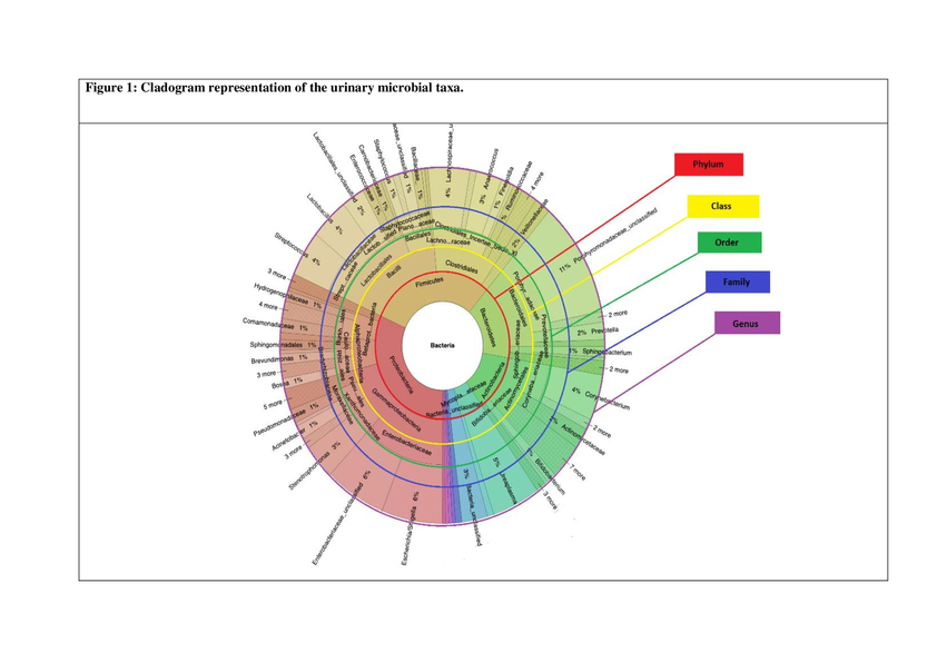
Objective: The aim of this study is establish the optimal non- invaszive urine sample collection method for the microbiota studies. Methodology: 12 men with bladder carcinoma underwent first voided and midstream urine collection. Urine samples were analyzed by using V3-V4 regions of bacterial 16s ribosomal RNAs. Bacterial groups with relative abundance above 1% were analyzed in first voided urine and midstream urine samples at phylum, class, order, and family level. At the genus level, all of the identified bacterial groups’ relative abundances were analyzed. The statistical significance (p<0.05) of differences between first voided and midstream urine sample microbiota were evaluated using the Wilcoxon test. Results: According to analysis, 8 phyla, 14 class, 23 orders, 39 families, and 29 different genera were identified in the first voided and the midstream urine samples. Statistical differences were not identified between first voided and mid-stream urine samples of all bacteria groups except the Clostridiales at order level (p:0.04) and Clostridia at class level (p:0.04). Conclusions: Either first voided or midstream urine samples can be used in urinary microbiota studies as we determined that there is no statistically significant difference between them regarding the results of 16s ribosomal RNA analysis. What’s known? According to widespread acceptance, first voided urine and midstream urine should be collected separately for standard microbiologic evaluation. What’s new? We found that there is no exact statistically significant difference between two collection methods even on microbiota analysis. We believe that either first voided or midstream uyrine samples can be used in urinary microbiota studies.