Ayse Demirtola

and 5 more

Dextrocardia is a cardiac positional anomaly in which the heart is located in right hemithorax with base-to-apex axis directed to the right and caudad. A number of congenital heart defects have been reported with dextrocardia, including VSD, PDA, ASD, TOF, pentalogy of Fallot, infundubular PS, transposition and pseudotruncus and total anomalous pulmonary venous return. We will share a patient with severe TR due to tricuspid valve commissural prolapse. A 42-year-old female patient was admitted to our clinic with the complaint of dyspnea. Transthoracic echocardiography revealed dextrocardia and severe TR consisting of two separate regurgitation jets. Contrast echocardiography performed due to dilatation of the coronary sinüs, did not show persistent left superior vena cava, and no right-to-left shunt was observed. Transesophageal echocardiography showed a prolapse in the commissure where the tricuspid anterior and septal leaflets meet, and a severe eccentric regurgitation jet with an area of vena contracta 0.75cm2 in the 3D MPR was observed. A moderate regurgitation jet was also seen from the coaptation line of all three leaflets. Commussural prolapse and regurgitation jet revealed in detail by 3D imaging. No significant pathology was detected in the other valves except mild insufficiency. Right heart catheterization and tricuspid valve surgery were planned for the patient with normal right heart functions. Although it is known that there are many congenital pathologies accompanying dextrocardia, we are happy to share our experience with you as the first case to report the coexistence of primary tricuspid valve disease and commusural prolapse with 3D detailed imaging.

Ayse Demirtola

and 5 more

Purpose: Cardiac resynchronization therapy (CRT) has a positive effect on the improvement of functional mitral regurgitation in patients with heart failure with reduced ejection fraction. However geometric changes in the mitral valve apparatus, subvalvular structures and their contribution to the improvement of mitral regurgitation after CRT have not been clearly defined. The aim of our study was to evaluate the geometric parameters of mitral valve apparatus measured with 3Dimensional (3D) transesophageal echocardiography (TEE) before CRT implantation and to determine the parameters predicting the improvement of mitral regurgitation after CRT. Methods: In this prospective study thirty patients with moderate or severe mitral regurgitation with low EF heart failure planned for CRT implantation and had an indication for TEE were included. Effective regurgitant orifice (ERO) and regurgitant volume (RV) measurements were performed before CRT implantation. Detailed quantitative measurements of mitral valve were done from recorded images by 3D TEE. ERO, RV measurements were repeated to evaluate mitral regurgitation at the end of 3rd month. Results: There were no significant changes in left ventricular EF and left ventricular diameters at third month follow-up, whereas ERO and RV values were decreased. posterior leaflet angle was found higher in non-responder group compared to responder group. (28,93 ± 8,41 vs 41,25 ± 10,90, p = 0,006). Conclusion: Heart failure patients with moderate or severe functional mitral regurgitation who underwent CRT implantation were found lower posterior leaflet angle which was measured by 3D TEE in the patient group whose mitral regurgitation improved after CRT.

Turkan Tan

and 12 more

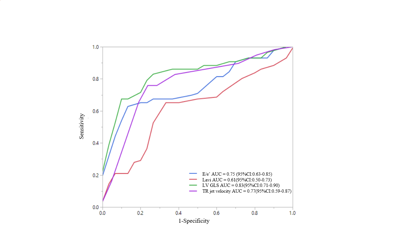
Purpose: An elevated left ventricular (LV) filling pressure is the main finding in patients with heart failure with preserved ejection fraction, which is estimated with an algorithm using echocardiographic parameters recommended by the recent American Society of Echocardiography (ASE)/European Association of Cardiovascular Imaging (EACVI) guideline. In this study, we sought to determine the efficacy of LV global longitudinal strain (GLS) to estimate the elevated LV filling pressure. Methods and Results: 73 prospectively selected patients undergoing left ventricular catheterization (mean age 63.19±9.64, 68.5% male) participated in this study. Using the algorithm, the LV filling pressure was estimated with the echo parameters obtained within 24hrs before the catheterization. The LV GLS was measured using the automated functional imaging system (GE, Vivid E9 USA). Invasive LV pre-A pressure corresponding to mean left atrial pressure (LAP) was used as a reference, and >12 mm Hg was defined as elevated. The invasive LV filling pressure was elevated in 43 (58.9%) and normal in 30 patients (41.1%). In 9 (12.3%) patients of 73 are defined as indeterminate based on the 2016 algorithm. Using the ROC method, -18.1% of LV GLS estimated the LV filling pressure (AUC=0.79, 73% specificity, 84% sensitivity) with higher sensitivity compared with the algorithm (AUC=0.76, 77% specificity, 72% sensitivity). Conclusions: We confirmed that the LV GLS is feasible and reproducible in estimating LV filling pressure. In addition, LV GLS highly predicts elevated LAP as E/e’ and TR jet velocity and may be used as major criteria for the diagnosis of HFpEF