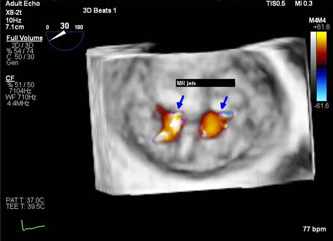
Case 1. An 82-year-old man with history of ischemic cardiomyopathy and multiple admissions due to acute decompensated heart failure was evaluated for moderate to severe secondary MR due to atrial dilation (atrial functional MR). TTE showed severe biatrial enlargement with a left atrial volume of 117mL and a left atrial volume index of 65.5ml/m2. It also showed LV of normal size, left ventricular LVIDd of 4.5cm and LVEF of 55%. En face view revealed two central jets arising from the coaptation gaps between posterior mitral leaflet indentations (P1/P2 and P2/P3) (Panel A). (Panel B) Transillumination rendering on 3D TEE, highlighted two distinct coaptation gaps between posterior mitral leaflet scallops. Case 2. A 63-years-old woman with medical history of ischemic cardiomyopathy and heart failure with reduced ejection fraction (35%) was evaluated for moderate to severe secondary MR. TTE showed the LV dilation with LVIDd of 5.7cm. TEE revealed severe eccentric MR. (Panel C) 3D color Doppler TEE imaging of the mitral valve showed a severe regurgitant jet, originated in-between P2 and P3 posterior scallops. (Panel D) Transillumination rendering on 3D TEE, view from left atrium, in systole highlighted the coaptation gap. (Panel E) 3D color Doppler TEE imaging showed residual mild MR after a mitral clip was deployed grasping the medial aspect of P2 and A2 scallops covering the coaptation defect. (Panel F) Transillumination rendering on 3D TEE, view from LV, showed complete resolution of the coaptation gap between posterior scallops after clip deployment.