Marcin Kuniewicz

and 1 more

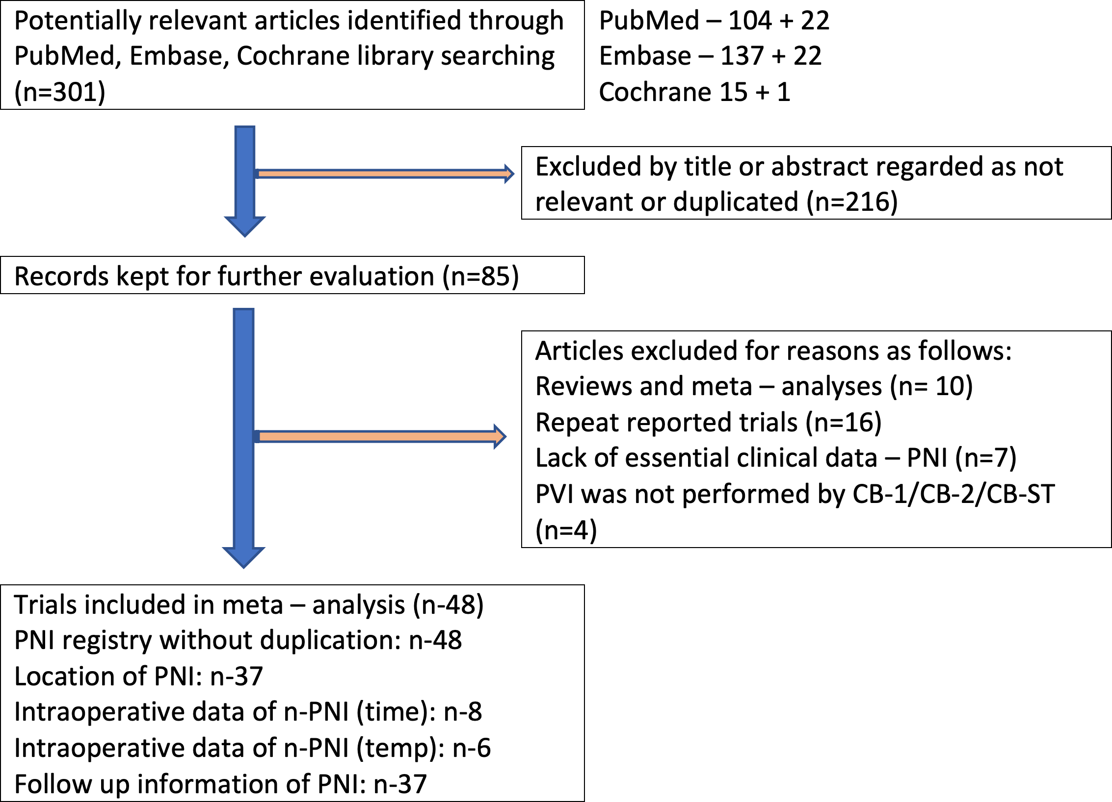
Background: The right phrenic nerve is vulnerable to injury (rPNI) during cryoballoon ablation (CBA) isolation of the right pulmonary veins. The complication can be transient or persistent. The reported incidence of rPNI fluctuates from 4.73% to 24.7% depending on changes over time, CBA-generation, and selected protective methods. Methods: Through September 2019, a database search was performed on MEDLINE, EMBASE, and Cochrane Database. In the selected articles, the references were also extensively searched. The study provides a comprehensive meta-analysis of the overall prevalence of rPNI, assesses the transient to persistent PNI-ratio, the outcome of using compound motor action potentials (CMAP), and estimated average time to nerve recovery. Results: From 2008 to 2019, 10,341 records from 48 trials were included. Out of 783 eighty- PNI (7.7%), 589 (5.7%) were transient, and 194 (1.9%) were persistent. CMAP caused a significant reduction in the risk of persistent PNI from 2.3% to 1.1% (p = 0.05; odds ratio [OR] 2.13) in all CBA groups. The mean time to rPNI recovery extended beyond the hospital discharge was significantly shorter in CMAP group at three months on average versus non CMAP at six months (p = 0.012). CMAP (in contrast to non-CMAP procedures) detects PNI earlier from 4 to 16 sec (p < 0.05; I2 = 74.53%) and 3 to 9ยบ (p < 0.05; I2 = 97.24%) earlier. Conclusions: rPNI extending beyond hospitalization is a relatively rare complication. CMAP use causes a significant decrease in the risk of prolonged injury and shortens the time to recovery

Marcin Kuniewicz

and 5 more

The left ventricular summit (LVS) is a triangular area located at the most superior portion of the left epicardial ventricular region, surrounded by the two branches of the left coronary artery: the left anterior interventricular artery and the left circumflex artery. The triangle is bounded by the apex, septal and mitral margins and base. This review aims to provide a systematic and comprehensive anatomical description and proper terminology in the LVS region that may facilitate exchanging information among anatomists and electrophysiologists, increasing knowledge of this cardiac region. We postulate that the most dominant septal perforator (not the first septal perforator) should characterize the LVS definition. Abundant epicardial adipose tissue overlying the LVS myocardium may affect arrhythmogenic processes and electrophysiological procedures within the LVS region. The LVS is divided into two clinically significant regions: accessible and inaccessible areas. Rich arterial and venous coronary vasculature and a relatively dense network of cardiac autonomic nerve fibers are present within the LVS boundaries. Although the approach to the LVS may be challenging, it can be executed indirectly using the surrounding structures. Delivery of the proper radiofrequency energy to the arrhythmia source, avoiding coronary artery damage at the same time, may be a challenge. Therefore, coronary angiography or cardiac computed tomography imaging is strongly recommended before any procedure within the LVS region. Further research on LVS morphology and physiology should increase the safety and effectiveness of invasive electrophysiological procedures performed within this region of the human heart.Published in Diagnostics:https://doi.org/10.3390/diagnostics11081423