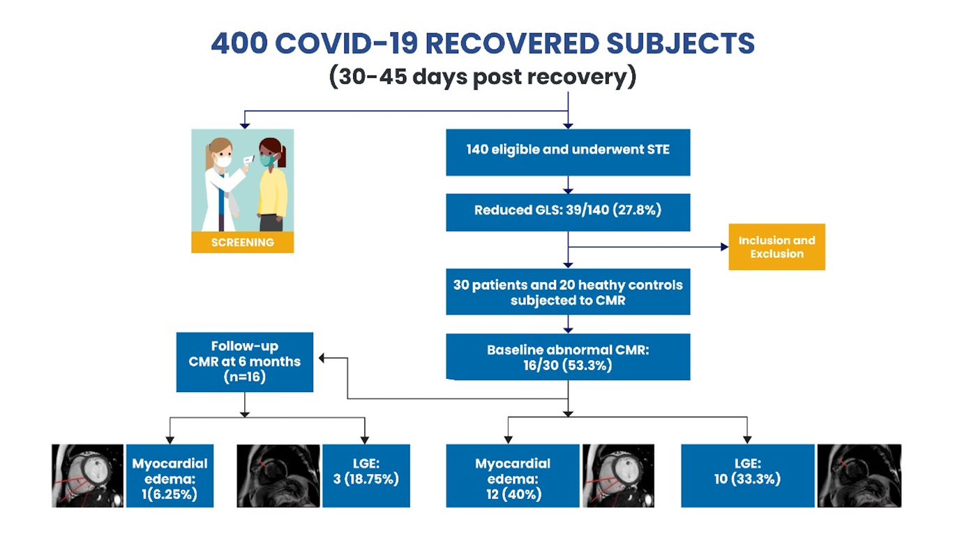
Objective: To evaluate for cardiac involvement in recovered COVID-19 patients using cardiac magnetic resonance imaging (MRI). Methods: A total of 30 subjects recently recovered from COVID-19 and abnormal left ventricular global longitudinal strain were enrolled. Routine investigations, inflammatory markers and cardiac MRI were done at baseline with follow-up scan at 6 months in individuals with abnormal baseline scan. Additionally, 20 age-and sex-matched individuals were enrolled as healthy controls (HCs). Results: All 30 enrolled subjects were symptomatic during active COVID-19 disease and were categorized as mild: 11 (36.7%), moderate: 6 (20%) and severe: 13 (43.3%). Of the 30 patients, 16 (53.3%) had abnormal CMR findings. Myocardial edema was reported in 12 (40%) patients while 10 (33.3%) had LGE. No difference was observed in terms of conventional LV parameters however, COVID-19 recovered patients had significantly lower right ventricular (RV) ejection fraction, RV stroke volume and RV cardiac index compared to HCs. Follow-up scan was abnormal in 4/16 (25%) with LGE persisting in 3 patients. Myocardial T1 (1284 + 43.8 ms vs 1147.6 + 68.4 ms; P<0.0001) and T2 values (50.8+16.7 ms vs 42.6+3.6 ms; P=0.04) were significantly higher in post COVID-19 subjects compared to HCs. Similarly, T1 and T2 values of severe COVID-19 patients were significantly higher compared to mild and moderate cases. Conclusions: An abnormal CMR was seen in half of recovered patients with persistent abnormality in one-fourth at six months. Our study suggests a need for closer follow-up among recovered subjects in order to evaluate for long term cardiovascular sequalae.