Utpal Gaikwad

and 7 more

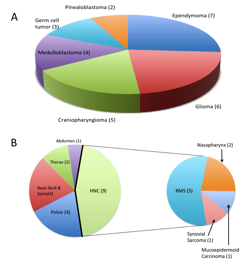
Background: To report our experience with image guided pencil beam proton beam therapy (PBT) for craniospinal irradiation (CSI). Materials and Methods: Between January 2019 to Dec 2021, we carried out a detailed audit of the first forty patients treated with PBT. We had recorded acute toxicities, reporting early outcomes and discuss limitations of current contouring guidelines during CSI PBT planning. Results: Median age of the patient cohort was 8 years, and histologies include 20 medulloblastoma, 7 recurrent ependymoma, 3 pineoblastoma, 3 were germ cell tumors and remaining 7 constituted other diagnoses. Forty percent patients received concurrent chemotherapy. Median CSI dose was 23.4 GyE (Gray Equivalent; range 21.6 - 35). Thirty-five patients (87.5%) completed their CSI without interruption, 5 required hospital admission. No patient had grade 2/> weight loss during the treatment. Forty-five percent (18) developed grade 1 haematological toxicities and 20% (8) developed grade 2 or 3 toxicities; none had grade 4 toxicities. At median follow up of 12 months, 90 % patients are alive of whom 88.9 % are having local control. Special consideration with modification in standard contouring used at our institute helped in limiting acute toxicities in paediatric CSI patients. Conclusion: Our preliminary experience with modern contemporary PBT using pencil beam technology and daily image guidance in a range of tumours suitable for CSI is encouraging. Patients tolerated the treatment well with acceptable acute toxicity and expected short-term survival outcome. In paediatric CSI patients, modification in standard contouring guidelines required to achieve better results with PBT.

Prerna Kartik

and 9 more

Background: The incidence of brain tumours in India equate to half of those in the developed world. Delayed diagnosis is associated with a higher risk of life-threatening neurological complications at presentation and poor cognitive outcomes amongst survivors. Early detection and treatment is crucial for improving outcomes. Aims: The aim of this study was to analyse baseline diagnostic intervals for paediatric brain tumours in Tamil Nadu. Methods: Data for this retrospective study was collected via questionnaire across 8 different hospitals in Tamil Nadu. It consisted of 14 questions where doctors were asked to record data items including the date of symptom onset, first presentation to healthcare and date of diagnosis. Results: 114 children were diagnosed with a brain tumour between January 2018 – October 2020. The average diagnostic interval was 9.3 weeks (median 3.5 weeks), and the average patient interval is 6.1 weeks (median 0.6 weeks.). Low-grade tumours had the longest median total diagnostic interval of 6.6 weeks. The median total diagnostic interval was significantly higher in villages (7.9 weeks), as compared to patients located in District (4.8 weeks) and cities (2.3 weeks). Conclusion: Overall, the diagnostic interval for paediatric brain tumours were comparable to data in the UK. Moreover, all patients received an MRI within a day, indicating excellent infrastructure.. However, many low-grade and optic pathway tumours were unaccounted for. Tamil Nadu has one of the best healthcare systems in India and extending this methodology to areas with poorer healthcare provisions, is required to get representative national data.

Srinivas Chilukuri

and 16 more

Background: We report demographic profile and our initial experience of treating children and young adults with image guided pencil beam scanning proton beam therapy (PBS-PBT) at our centre. Material and methods: All patients younger than 25 years, consecutively treated with PBT based on a multi-disciplinary tumor board decision were analyzed. Patients were treated under daily on-board kilovoltage x-ray and/or cone beam CT scan guidance. The demographic profile, treatment characteristics and the acute toxicities were reported. Patient and treatment related factors and their association with acute toxicities were analyzed using univariate and multivariate analysis. Results: Forty-seven patients {27 with central nervous system(CNS) and 20 with non-CNS tumors} with a median age of 9 years were evaluated. Most common diagnoses were ependymoma, rhabdomyosarcoma and glioma. Median dose delivered was 54.8CGE(40-70.4) to a median clinical target volume of 175cc (18.7-3083cc) with 34% requiring concurrent chemotherapy(CCT). Acute grade-2 and 3 dermatitis, mucositis, and hematological toxicity was noted in 45% and 2%; 34% and 0%; 38% and 30%; respectively. Grade-2 fatigue was noted in 26%. On univariate analysis, CCT(p=0.009) and cranio-spinal irradiation(p<0.001) were associated with grade-2 or more hematological toxicity in patients with CNS tumors. Among non-CNS tumors, clinical target volume more than 150cc was associated with grade-2 or more fatigue(p=0.017). Conclusions: The demographic pattern of patients treated with PBT at this new and only centre in the region was similar to previously published literature. Image guided PBS-PBT resulted in acceptable acute toxicities both among children with CNS and non-CNS tumors.