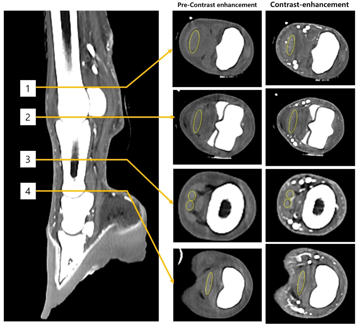
Background: Few studies have investigated the adequate contrast enhancement (CE) evaluation depending on concentration, volume, and rate of contrast media (CM) and the scan parameters in equine contrast enhanced computed tomography (CECT). Objectives: We investigated CE of the deep digital flexor tendon (DDFT) and arteries depending on voltage, concentration, volume, and rate of CM during intra-arterial CECT of equine distal forelimbs. Study design: This is a prospective study. Methods: Six horses underwent fifty-four CT scans. First, the CE of DDFT and arteries was evaluated depending on the voltage (80 or 120 kV) and CM concentration (150, 120, or 90 mg I/mL in 50 mL of CM). Second, CE of DDFT and vessels was evaluated depending on the CM volume (50, 100, or 150 mL) and administration rate (2, 4, or 6 mL/s) with a fixed iodine delivery rate (IDR) (300 or 180 mg I/s). Results: CE of DDFT significantly increased at 80 kV of voltage and 150 mg I/mL of CM concentration (Median: 29.65; IQR: 1.74; P < 0.05). CE of the DDFT positively correlated with CM concentration (P = 0.0004; r = 0.75). At 180 mg I/s of IDR, an increase in rate and volume (6 mL/s and 150 mL) led to low contrast attenuation in the medial and lateral palmar arteries (median and IQR: 985.93 and 71.8 Hounsfield units [HU] and 988.73 and 41.16 HU, respectively); the CE was sufficient to distinguish the artery from the adjacent structures. Main limitations: The number of animals was small for parametric statistical analysis. Conclusions: Our results suggest that a low CM concentration could yield sufficient CE of the DDFT and arteries with adjusted CT scanning parameters or volume and injection rate of CM.