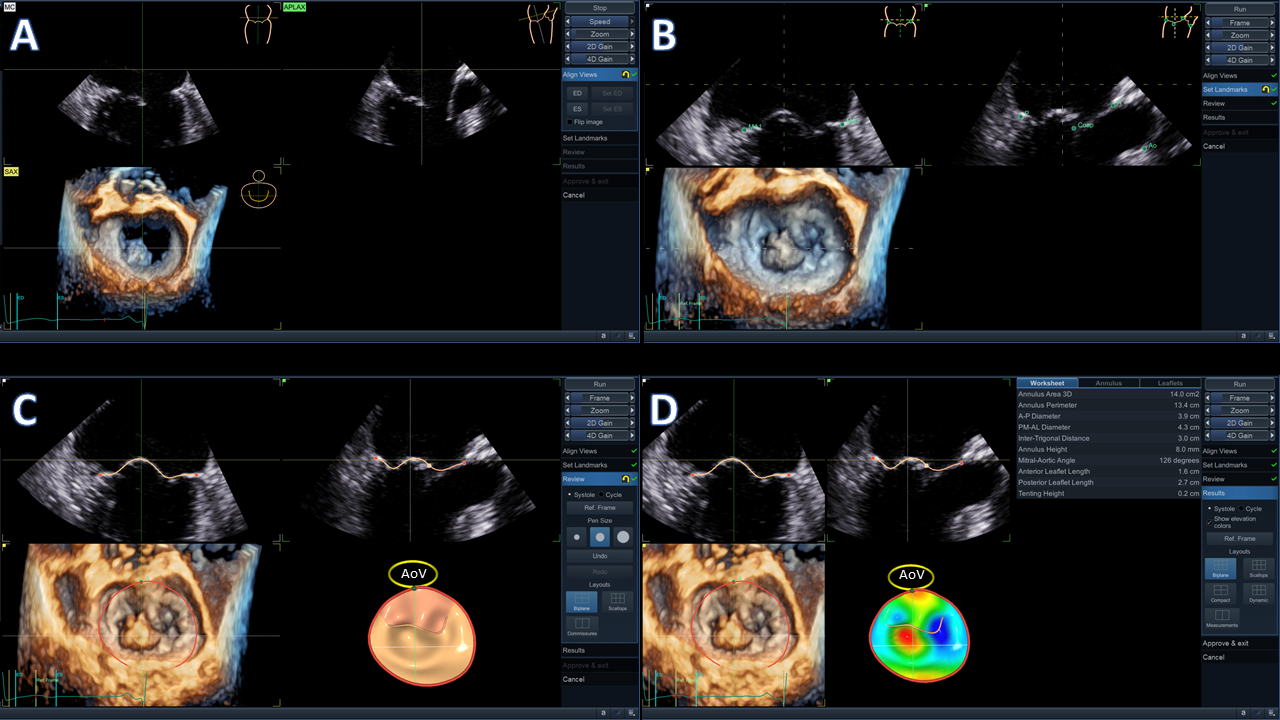
Discrimination of myocardial function changes: evolution of chronic kidney disease or hemodialysis effect?Kalliopi Keramida, MD, MSc, PHD, FESC, FHFA, FICOS,1Konstantinos Papadopoulos, MD, PhD, FESC, FEACVI21 Cardiology Department, General Anti-Cancer, Oncological Hospital Agios Savvas, Athens, Greece2Echocardiography department, Interbalkan Medical Center, Thessaloniki, GreeceCorresponding author: Kalliopi Keramida, General Anti-Cancer Oncological Hospital, Agios Savvas Athens, GreeceAleksandras Avenue 171, 11522, Athens, Greece,[email protected] on “Effect of hemodialysis on left atrial function in patients with end-stage renal failure evaluated by two-dimensional speckle-tracking imaging”Chronic kidney disease, often advancing to end-stage renal disease (ESRD), poses a significant challenge to public health, as costly treatments like dialysis or kidney transplantation become essential for survival. Globally, the number of individuals with ESRD that need renal replacement therapy is estimated to range between 4.902 and 9.701 million with a trend to double by 2030 (1). Cardiovascular (CV) disease is a prevalent complication and the leading cause of mortality among individuals with ESRD undergoing hemodialysis (HD). Heart failure (HF), sudden cardiac death, ischemic cardiomyopathy and stroke are the main causes of CV mortality (2). Echocardiography is the main imaging modality used for the long-term surveillance of ESRD patients.Myocardial remodeling and dysfunction, are prevalent in different stages of chronic kidney disease (CKD), leading to uremic cardiomyopathy and HF with either preserved (3) or reduced ejection fraction later in the course of the disease (4). However, myocardial remodeling, including left ventricular (LV) hypertrophy, dilatation, fibrosis, and dysfunction are not confined to the LV only. The left atrium (LA) is also affected by this remodeling process. LA dilatation and dysfunction have been studied in several trials, revealing their prognostic value in patients with CKD (5, 6). Miao Y et colleagues (7) used two-dimensional speckle tracking imaging to assess concurrently LA and LV function changes induced by HD in ESRD patients. The authors demonstrated LV and LA remodeling in ESRD patients compared to controls (increased LV wall thickness, LV volumes and LA volume), with higher rates of diastolic dysfunction and elevated filling pressures measured by E/E’ ratio, but LV systolic performance, assessed by LV ejection fraction (LVEF) and LV Global Longitudinal Strain (LVGLS) similar to controls. Further assessment of LA function indices [reservoir (LASr), conduit (LAScd) and contractile (LASct) strain], demonstrated significantly baseline reduced LA strain values, compared to controls, and further reduction of LASr and LAScd after HD. LASct remained stable without further deterioration after HD (7).This elegant study included a control group to compare the left heart strain values between patients with ESRD and healthy subjects and delineated the undeniable remodeling of both left heart chambers (7). Although the baseline echocardiographic parameters used for LV systolic function assessment (LVEF, LVGLS) did not differ between controls and ESRD patients in this study, previously published trials have shown that ESRD patients have impaired LVGLS (8) and myocardial work indices (9, 10). Furthermore, there is consensus in literature, that these patients have diastolic dysfunction expressed by increased LA volume and E/e’, findings relevant to the LV remodeling with wall thickening and increased filling pressures. A more sensitive marker of diastolic dysfunction in HF, irrespective of LVEF, is the LA strain (11) that has already proven its prognostic role in ESRD patients (6). However, clinicians and researchers need to be careful, as the three different LA strain parameters, reflect different properties of LA and LV and are related to volume status. More specifically, LASr expresses LA pressure and/or LA stiffness. LAScd reflects LV relaxation and/or LV pressure in early diastole, while LASct is indicative of LV compliance, LV end-diastolic pressure and/or LA contractility (5). LA strain is load-dependent and previous research has demonstrated that it is more accurate in depressed EF rather than in normal and if we are about to be specific for increased LV filling pressures, we need to use the LASr<18% as a cut-off value (12).The changes induced by HD in strain-related parameters of LV and LA, are explained by their pre- and after-load dependency. LVGLS was decreased after HD due to decrease of preload and afterload (decreased systolic blood pressure), decrease of LV end-diastolic volume and myocardial stunning and troponin release (13). The inclusion of myocardial work (MW) analysis in the echocardiographic assessment of these patients, can help in the clarification of the cause of LVGLS change after HD (9). Myocardial work includes the afterload of the patient by measuring the blood pressure with a simple brachial cuff and it is less-load dependent than GLS. A meta-analysis for LV strain evaluation showed reduction of LV GLS when afterload is increased, which may lead to false conclusions about the LV performance (14). In HD patients, reduction of both LVGLS and MW would suggest true deterioration of systolic performance, while reduction of GLS with increase of MW would detect blood pressure changes and not LV systolic impairment.LASr and LAScd components are decreased immediately after HD, confirming their load dependency, while LASct remains unchanged, being independent of volume status and preload (7). LASct mainly reflects LA systolic function and LV compliance (7), related to the disease progression. The effects of blood pressure changes induced by HD, are not included in the assessment of LA function. A less-load dependent marker of LA function, maybe similar to LV MW (namely “LA Myocardial Work”), taking into account volume and pressure changes, would be more sensitive in detecting acute LA cardiomyopathy and diastolic dysfunction in ESRD patients after HD. Research should be done in that way, including large series of healthy subjects first and patients as well with different stages of diastolic and systolic dysfunction. If such marker is developed, this will detect definitely atrial myopathy in different populations with applicability in CKD patients. The current application of MW is validated for LV function and with afterload involvement only (15). No preload data or increased LV diastolic pressures are included into the equation. This method looks promising for accurate evaluation of the true LV work, but more research should be done in different populations.In concluding, LV GLS and LA strain are sensitive markers that can detect subclinical LV dysfunction and atrial myopathy but with the restrictions of load and pressure dependency. Myocardial Work may further help discriminate the true impairment of the LV function in ESRD patients and can be included into the echocardiographic follow-up protocol of these patients (Figure). A new marker of LA function, independent from volume changes and LV filling pressures should be developed in order to detect subtle changes of LA function and possibly guide the treatment in this high-risk population.