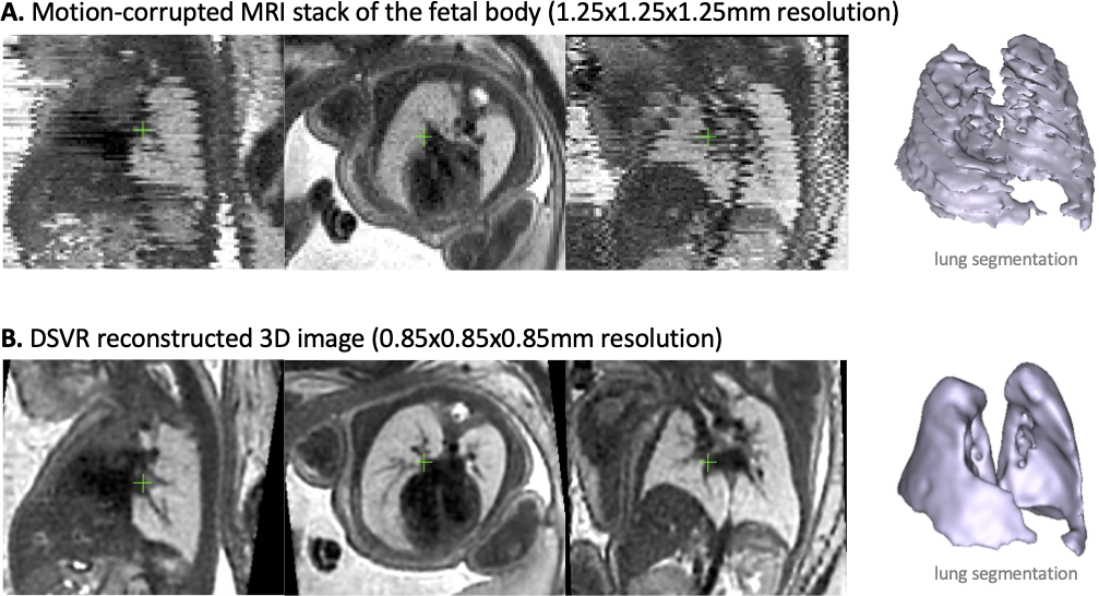
Abstract Objective:Evaluate deformable slice-to-volume registration (DSVR) to calculate 3D-segmented total lung volume (TLV) in fetuses with congenital diaphragmatic hernia, congenital lung lesions and healthy controls, with comparison to 2D-manual segmentation. Design:Pilot study Setting:Regional fetal medicine referral centre Sample:Fetal MRIs performed for clinical indications (abnormal cases) or as research participants (healthy controls) Methods:Sixteen MRI datasets of fetuses (22-32 weeks GA). Diagnosis: CDH(n=5), CPAM(n=2), CDH with BPS(n=1) and healthy control(n=8). DSVR was used for reconstruction of 3D isotropic (0.85 mm) volumes of fetal body followed by semi-automated lung segmentation. The resulting 3D TLV were compared to the traditional 2D-based volumetry, and a normogram of DSVR-derived fetal lung volumes from 100 cases was produced. Main Outcome Measures:Concordance with 2D-volumetry assessed with Bland-Altman analysis, results of segmentations presented visually. Observed/Expected values were calculated for abnormal cases based upon the normogram. Results:DSVR-derived TLV values have high correlation with the 2D-based measurements but with a consistently lower volume; bias -1.44cm3 [95% limits: -2.6 to -0.3] with improved resolution able to exclude hilar structures even in severe motion corruption or in cases of lung hypoplasia. Conclusions:Application of DSVR for fetal MRI provides a solution for analysis of motion corrupted scans and does not suffer from the interpolation error inherent in 2D-segmentation as per current clinical practice. It increases information content of acquired data in terms of visualising organs in 3D space and quantification of volumes, which we believe will have important value for counselling and surgical planning. Keywords:Fetal MRI; congenital diaphragmatic hernia; CPAM; lung volume