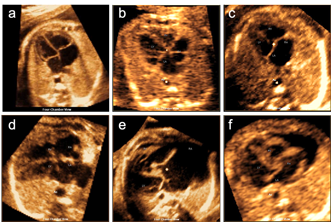
Attempting a comprehensive examination of the fetal heart remains challenging for unexperienced operators as it emphasizes the acquisition and documentation of sequential cross-sectional and sagittal views and inevitably results in diminished detection rates of fetuses affected by congenital heart disease. The introduction of three-/four-dimensional spatial-temporal image correlation 3D/4D STIC technology facilitated a volumetric approach for thorough cardiac anatomic evaluation by the acquisition of cardiac 4D datasets by analyzing and correlating numerous images from different heart cycles obtained during an automated sweep and subsequently displaying them in an endless cine loop sequence. However, postanalysis with manipulation and repeated slicing of the volume usually requires experience and in-depth anatomic knowledge, which limits the widespread application of this advanced technique in clinical care and unfortunately leads to the underestimation of its diagnostic value to date. Fetal intelligent navigation echocardiography (FINE), a novel method that automatically generates and displays 9 standard fetal echocardiographic views in normal hearts, has shown to be able to overcome these limitations. Very recent data on the detection of congenital heart defects (CHDs) revealed a sensitivity and specificity of 98 % and 93 %, respectively. In this two-part manuscript, we focused on the performance of FINE in delineating abnormal anatomy of typical right and left heart lesions and thereby emphasized the educational potential of this technology for more than just teaching purposes. We further discussed recent findings regarding these morphological changes seen in a pathophysiological and/or functional context.