Jakub Holda

and 8 more

Introduction: The aim of this study was to describe the morphology of the right atrioventricular valve (RAV) and determine its spatial position in relation to selected structures of the right atrium. Methods and Results: We examined 200 randomly selected human adult hearts. All leaflets and commissures were identified and measured. The position of the RAV was defined. 3-leaflet configurations were present in 67.0% of cases whereas 4-leaflet configurations were present in 33.0%. Valves with four leaflets have significantly larger perimeter (119.2±11.1 vs. 109.3±11.3mm, p=0.001). No significant difference was found in superior leaflet length and height between 3- and 4-leaflet RAVs. Septal and mural leaflets were both significantly shorter and higher in 4-leaflet than in 3-leaflet RAVs. Significant domination of the muro-septal commissure in 3-leflet valves was noted. The supero-septal commissure was the most stable point within RAV circumference, with no difference in its position between 3- and 4-leaflet valves. In 3-leaflet valves the muro-septal commissure was placed within cavo-tricuspid isthmus area in 52.2% of cases, followed by the right atrial appendage vestibule region (20.9%). In 4-leaflet RAVs, the infero-septal commissure was located predominantly in the cavo-tricuspid isthmus area and infero-mural commissure was always located within the right atrial appendage vestibule region. Conclusions: The RAV is a highly variable structure. The supero-septal part of the RAV is the least variable component, whereas the infero-mural is the most variable. The number of detected RAV leaflets significantly influences the relative position of individual valve components in relation to right atrial structures.

Marcin Kuniewicz

and 5 more

The left ventricular summit (LVS) is a triangular area located at the most superior portion of the left epicardial ventricular region, surrounded by the two branches of the left coronary artery: the left anterior interventricular artery and the left circumflex artery. The triangle is bounded by the apex, septal and mitral margins and base. This review aims to provide a systematic and comprehensive anatomical description and proper terminology in the LVS region that may facilitate exchanging information among anatomists and electrophysiologists, increasing knowledge of this cardiac region. We postulate that the most dominant septal perforator (not the first septal perforator) should characterize the LVS definition. Abundant epicardial adipose tissue overlying the LVS myocardium may affect arrhythmogenic processes and electrophysiological procedures within the LVS region. The LVS is divided into two clinically significant regions: accessible and inaccessible areas. Rich arterial and venous coronary vasculature and a relatively dense network of cardiac autonomic nerve fibers are present within the LVS boundaries. Although the approach to the LVS may be challenging, it can be executed indirectly using the surrounding structures. Delivery of the proper radiofrequency energy to the arrhythmia source, avoiding coronary artery damage at the same time, may be a challenge. Therefore, coronary angiography or cardiac computed tomography imaging is strongly recommended before any procedure within the LVS region. Further research on LVS morphology and physiology should increase the safety and effectiveness of invasive electrophysiological procedures performed within this region of the human heart.Published in Diagnostics:https://doi.org/10.3390/diagnostics11081423

Jakub Holda

and 7 more

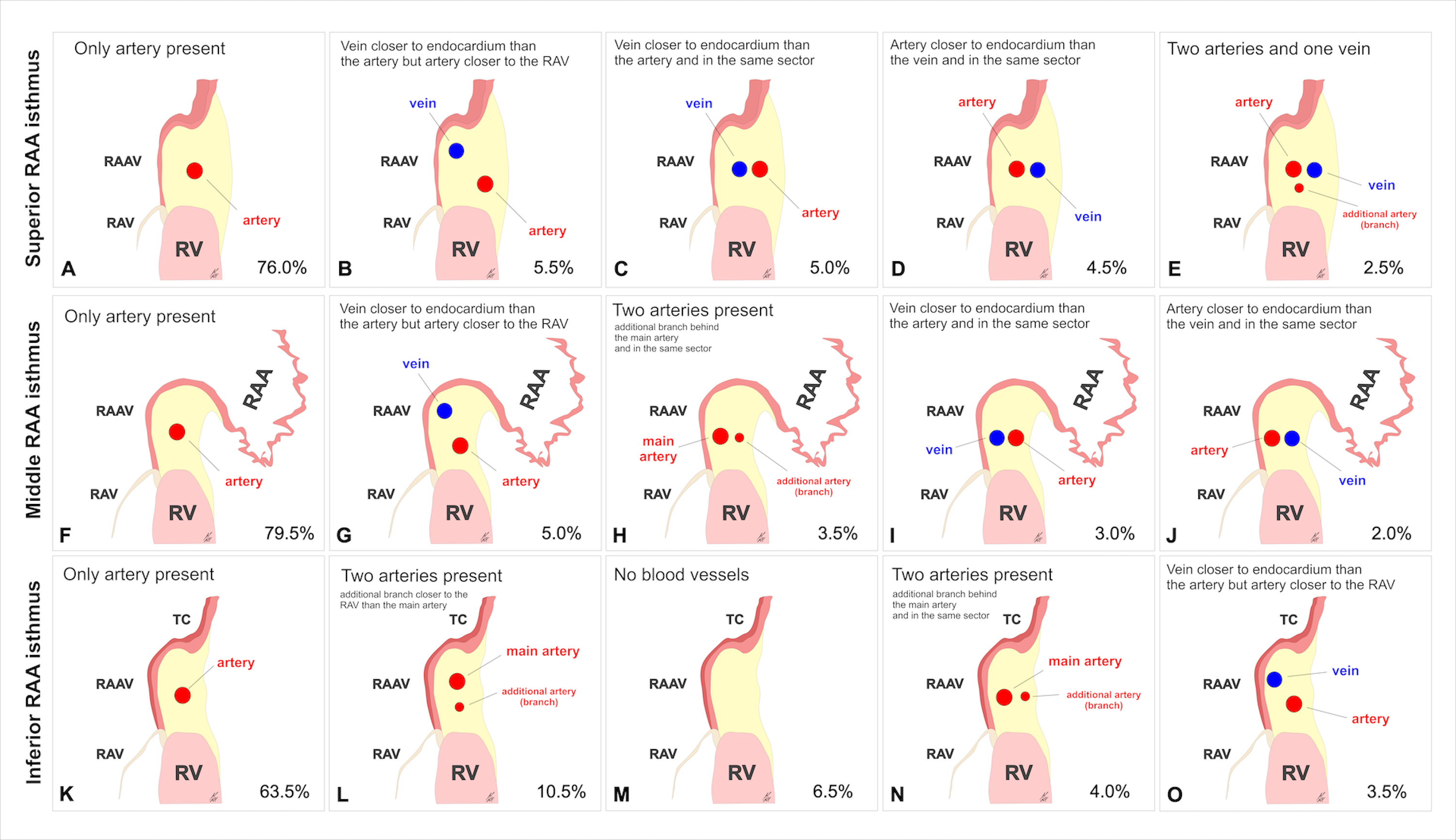
Introduction: The aim of our study was to investigate the presence and mutual relationships of coronary vessels within the right atrial appendage RAA vestibule. Methods and Results: We examined 200 autopsied hearts. The RAA vestibule was cross sectioned along its isthmuses (superior, middle, and inferior). We assessed the presence and mutual relationships between coronary blood vessels. The right coronary artery (RCA) was present in 100% of the superior RAA isthmuses but absent in 2.0% of hearts within the middle isthmus and in 6.5% of hearts within the inferior RAA isthmus. Its diameter was quite uniform along the superior (2.6±0.8mm), middle (2.9±1.1mm) and inferior (2.7±0.9mm) isthmuses (p=0.12). The location of the RCA varied significantly, and it was sometimes accompanied by other accessory coronary vessels. In all the isthmuses, the RCA ran significantly closer to the endocardial surface than to the epicardial surface (p<0.001). At the superior RAA isthmus, the artery was furthest from the right atrial endocardial surface and this distance gradually decreased between the middle RAA isthmus and the inferior RAA isthmus (9.0±4.0 vs. 6.2±3.0 vs. 4.8±2.3mm, respectively; p<0.001). The interposed RCA was found in 7.0% of cases within the superior isthmus, in 2.5% within the middle isthmus and in 1.5% within the inferior isthmus. Conclusions: This study was the most complex analysis of the mutual arrangements and morphometric characteristics of coronary blood vessels within the RAA vestibule. Awareness of additional blood vessels within the vestibule can help clinicians plan and perform safe and efficacious procedures in this region.