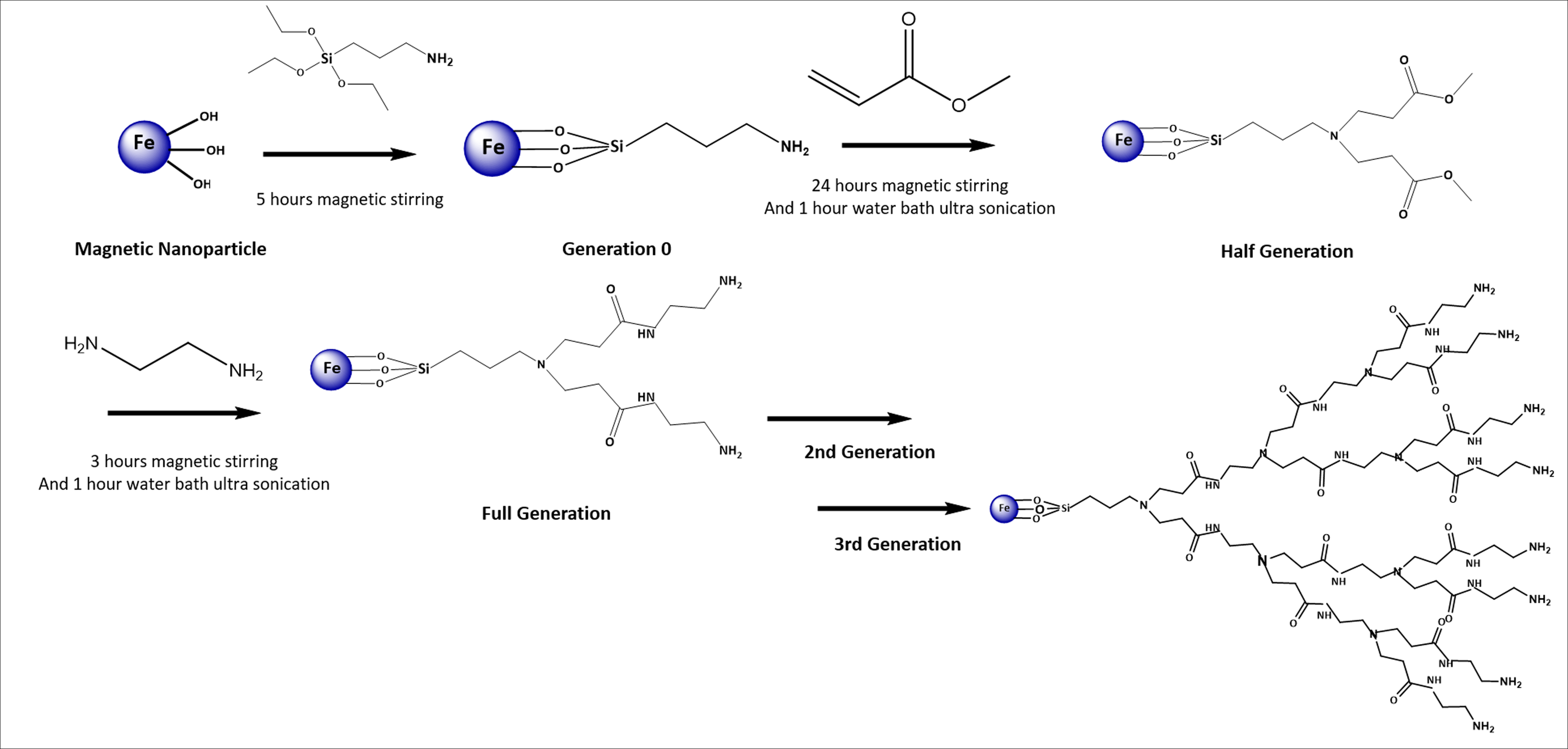
Nanotechnology plays a promising role in biomedical applications, particularly tissue engineering. Recently, the application of magnetic scaffolds and pulsed electromagnetic field (PEMF) exposure has been considered in bone tissue regeneration. In this study, 3rd generation dendrimer-modified superparamagnetic iron oxide nanoparticles (G3-SPIONs) are synthesized comprehensively characterized. Magnetic polycaprolactone (PCL) nanofibers are prepared by incorporating G3-SPIONs within the electrospinning process ,and physicochemical characteristics ,as well as cytocompatibility and cell attachment are assessed. Eventually, the osteogenic differentiation ability of adipocyte-derived mesenchymal stem cells (ADMSCs) cultured on the magnetic scaffold with and without PEMF exposure was investigated by measurement of alkaline phosphatase (ALP) activity and calcium content. The expression of specific bone markers was studied using the Real-time PCR method. According to the results, G3-SPIONs with mean size and zeta potential of 17.95 ± 3.57 nm and 22.7 mV, respectively, show a high saturation magnetization (57.75 emu/g). Adding G3-SPIONs to PCL significantly decrease nanofibers size to 495±144 nm and improves cell attachment and growth. The ADMSCs cultured on the G3-SPION-PCL scaffold in the presence of osteogenic media (OM) and exposure to PEMF expressed the highest Osteocalcin and Runx2 and showed higher calcium content as well as ALP activity. It can be concluded that the synthesized G3-SPION incorporated PCL nanofibers serve as a promising magnetic scaffold for bone regeneration. Also, utilizing the magnetic scaffold in the presence of OM and PEMF provides a synergistic effect toward osteogenic differentiation of ADMSCs. Key Words: Superparamagnetic iron oxide nanoparticles, Dendrimer, Polycaprolactone, Pulsed electromagnetic field, Bone tissue engineering