Longhai TIAN

and 3 more

A manifest nodoventricular accessory pathway with unusual electrophysiological manifestationsLonghai Tian MM,Long YANG MD, Yidong ZHAO MM,Qifang Liu MDDepartment of Cardiology,Guizhou Provincial People’s Hospital, Guiyang , ChinaCorresponding author: Qifang Liu E-mail:liuxu19782000 @163.comDisclosures:(None)Funding:This study was supported by Clinical Research Center Project of Department of science and technology of Guizhou Province.Key Words: Nodoventricular pathway, Preexcitation, Decremental conductionAbstractWe present an unusual case of a nodoventricular accessory pathway showing the degree of anterograde decremental conduction was more than the atrioventricular node decremental Conduction. In the patient,there was no rapid antegrade conduction and inducible reentrant tachycardias during electrophysiologic examine and no ablation was performed.Case reportA 24-year-old male was diagnosed to have preexcitation suggestive of a septal accessory pathway by a 12-lead ECG (Figure 1). Given his occupation, he was referred to our hospital for electrophysiologic testing and radiofrequency catheter ablation. During sinus rhythm, intracardiac electrograms revealed preexcitation with an A-H interval 80ms and H-V interval of 20ms. Ventricular overdrive pacing at a cycle length of 400ms resulted in ventriculoatrial dissociation. Atrial extra-stimuli (AES) were introduced during sinus rhythm with a cycle length of 500 ms, The coupling interval was reduced from 400 to 280 ms. This resulted in a gradual prolongation of the atrio-His interval that was associated with a prolongation of the H-V interval and a decrease in the degree of preexcitation (figure2). A decremental atrioventricular pathway was noted. Interestingly, This electrophysiologic phenomenon of decremental conduction suggests that the degree of anterograde decremental conduction of accessory pathway was more than the atrioventricular node (AVN).Atrial overdrive pacing (AOD) resulted in intermittent loss of preexcitation, PR prolongation, and Mobitz II atrioventricular block. At atrial pacing with cycle length of 300 ms, the H-V interval was suddenly increased to 102 ms and the QRS complex morphology showed a right bundle branch block pattern. This suggests anterograde conduction over accessory pathway and the QRS complex morphology presenting fully preexcitation (figure3). The ventricular insertion site of accessory pathway could have been predicted by the fully pre-excited QRS complex morphology. Spontaneous junctional beat was seen during electrophysiologic study. Consequently, the H-V interval was still 28ms and the QRS complex morphology remained minimal preexcitation,indicating the proximal insertion site of accessory pathway is AV node (figure 2).DISCUSSIONNodoventricular pathways(NVP) are rare accessory pathway that connect the posterior extensions of the AVN to the crest of the ventricular septum [1]. These pathways were anatomically described in 1941 by Mahaim and Winston. The diagnosis of manifest NVP is based on the following evidence: (1) upper insertion site is AVN, (2) lower insertion site is ventricular, (3) proving accessory pathway presence with decremental conduction manifestations and excluding fasciculoventricular pathway.In our case,the patient present preexcitation without symptoms of arrhythmias. The electrophysiologic study demonstrated evidence of anterograde decremental conduction in response to programmed atrial pacing. The QRS morphology and H-V intervals remained changed during AES, excluding a fasciculoventricular fiber. If the pathway was fasciculoventricular, preexcitation should remained fixed and H-V remained constant when conducting over either fast or slow pathways. Catheter pressure over AVN region resulted in junctional ectopy that remained preexcitation, indicating the proximal insertion site of accessory pathway is AV node. The sudden appearance of infra-His conduction block by AOD resulted in complete ventricular preexcitation,we can confirmed the distal insertion site is the septal of left ventricular based on fully pre-excited QRS morphology. All these findings indicate the presence of a manifest NVP pathway in the patient.In this case, on programmed atrial stimulation testing, there was decremental conduction over the pathway, resulting in a longer A–V and H-V time as the A1–A2 coupling interval decreased. As far as we are aware, this case is unique since the nodoventricular fiber show the degree of anterograde decremental conduction was more than the atrioventricular node. In contrast, most of the NVP bypass only a portion of the AV node with decremental conduction properties. Therefore, the preexcitation degree may increase and the H-V interval may decrease even to negative values during AES [2]. As far as our patient is concerned,The electrophysiologic features of anterograde decremental conduction may be due to the anisotropic conduction of the transitional area of the AV node and variability in the space construction of tissue[3]. In addition, the differential expression of connexin isoforms in the nodal area also may be responsible for more degree decremental conduction of NVP than AVN[4].[1] Nazer B, Walters TE, Dewland TA, et al. Variable Presentations and Ablation Sites for Manifest Nodoventricular/Nodofascicular Fibers. Circ Arrhythm Electrophysiol. 2019;12(9):e007337.[2] Ali H, Sorgente A, Lupo P, et al. Nodo- and fasciculoventricular pathways: Electrophysiological features and a proposed diagnostic algorithm for preexcitation variants. Heart Rhythm. 2015 ;12(7):1677-1682.[3] Katritsis DG, Marine JE, Latchamsetty R, et al. Coexistent Types of Atrioventricular Nodal Re-Entrant Tachycardia: Implications for the Tachycardia Circuit. Circ Arrhythm Electrophysiol. 2015;8(5):1189-1193.[4] Anderson RH, Quintana DS, Mori S, et al. Re-evaluation of the structure of the atrioventricular node and its connections with the atrium. Europace. 2020 ;22(5):821-830.Figure 1:Twelve-lead electrocardiographic recordings showing fixed and minimal preexcitation during sinus rhythm.Figure 2:Intracardiac recordings during programmed atrial stimulation demonstrating decreased preexcitation with a prolongation of the H-V interval. The last spontaneous junctional beat still showing a preexcitation morphology. Note that the QRS complex morphology change of the lead I and V1. His electrograms are recorded on the RV pole and red arrows indicate His potentials.Figure 3:Intracardiac recordings during atrial overdrive stimulation, demonstrating a longer HV interval and QRS complex morphology with fully preexcitation. Red arrows indicate His potentials.

LIU Qifang

and 3 more

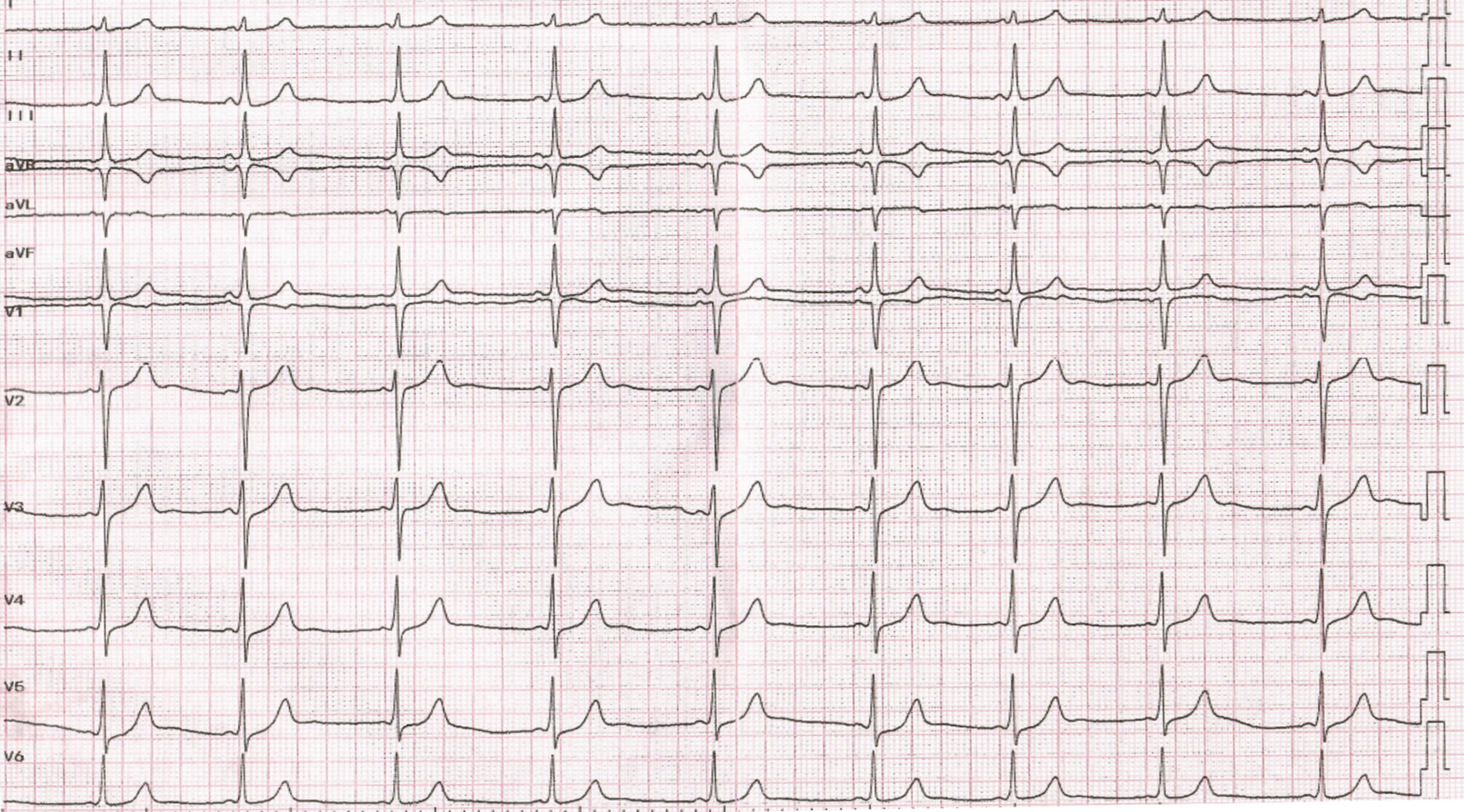
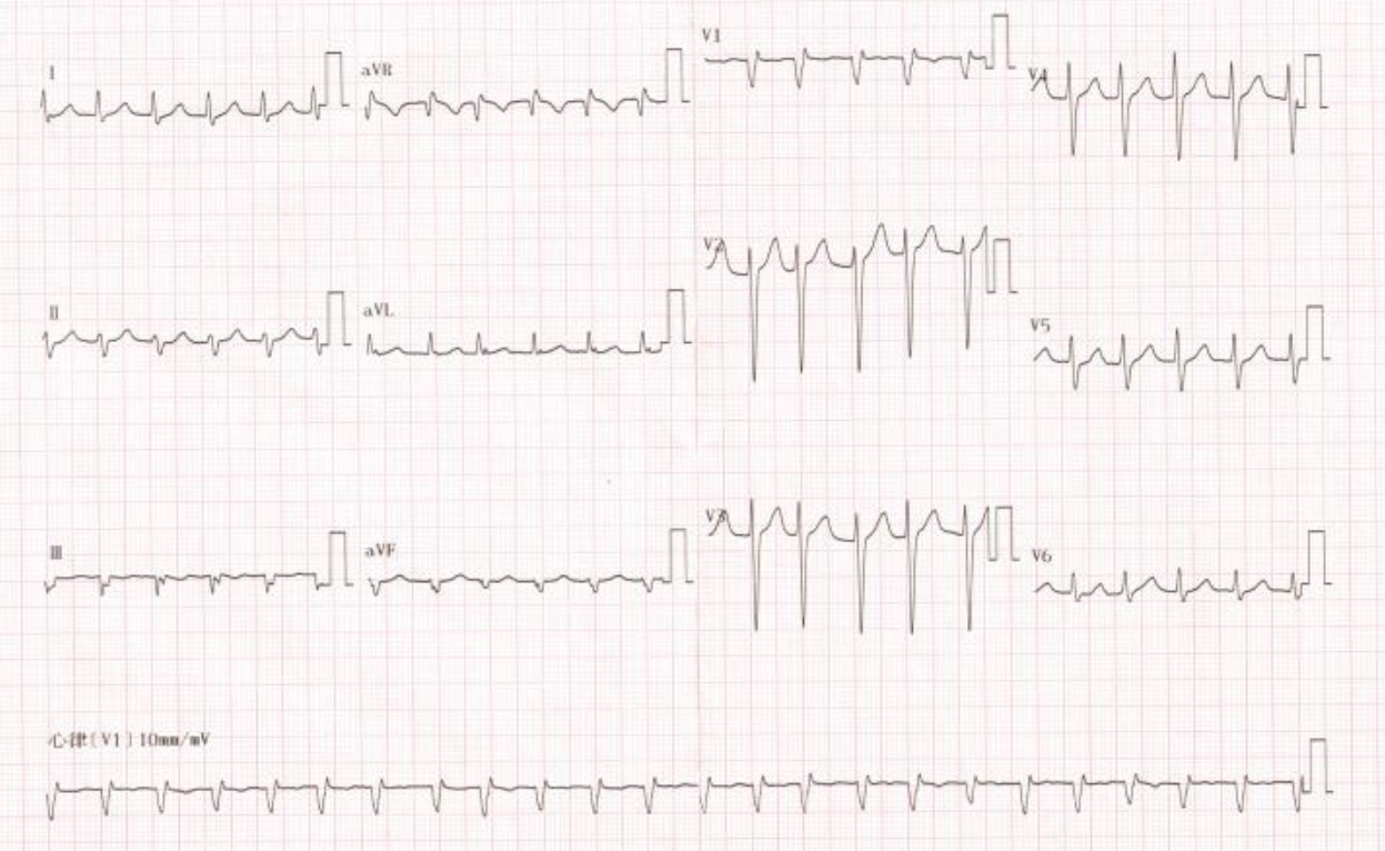
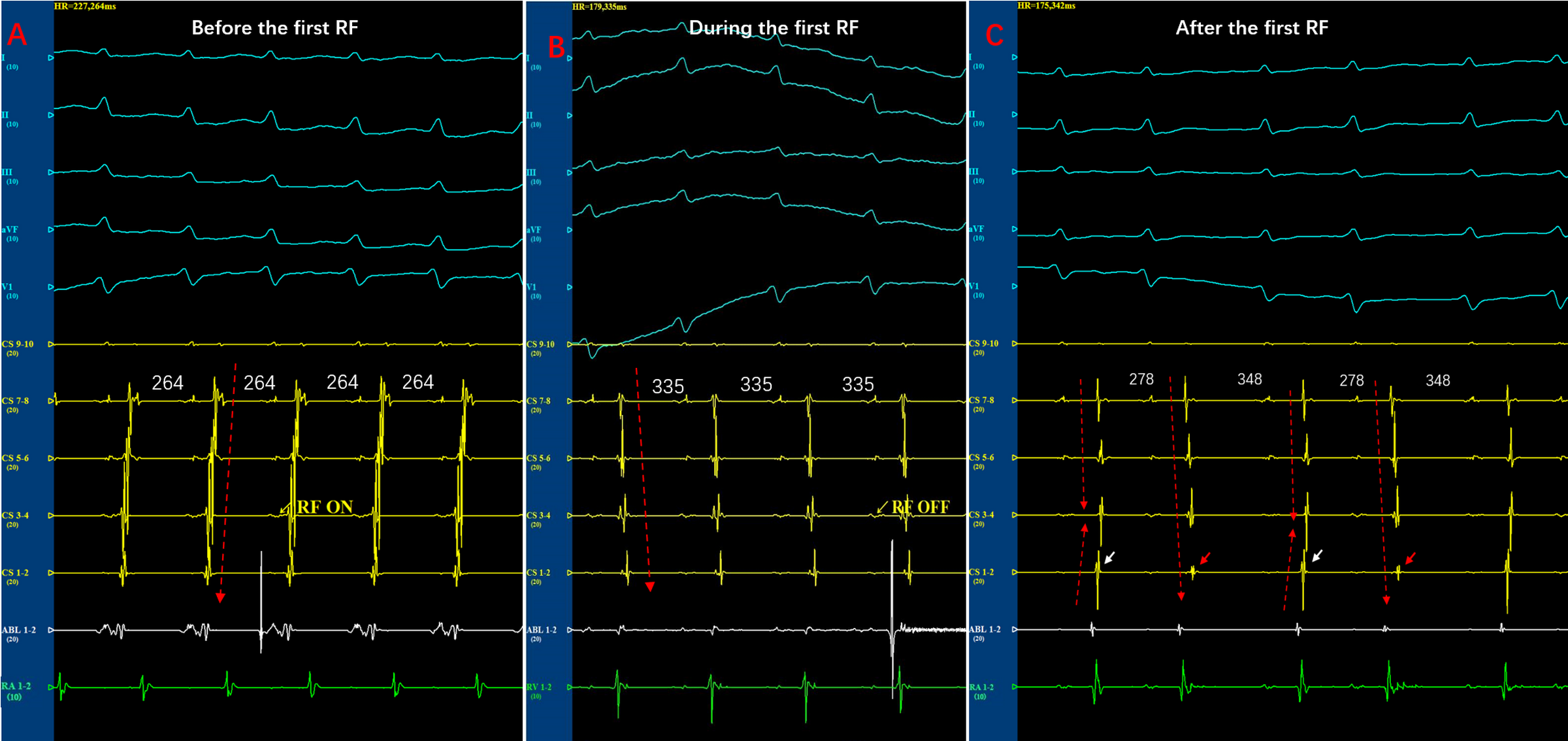
A narrow QRS complex tachycardia with irregular rhythm: what Is the mechanism?Liu Qifang MD, Tian Ye MD, Zhao Yidong MM, Yang Long MD.PHD,Department of Cardiology,Guizhou Provincial People’s Hospital, Guiyang , ChinaCorresponding anthor: Yang Long E-mail:[email protected]:(None)Funding: Clinical Research Center Project of Department of science and technology of guizhou province [NO. (2017)5405]Key Words: Atrioventricular nodal reentrant tachycardia;Catheter ablation;Irregular rhythmAbstractIn most patients with atrioventricular nodal reentrant tachycardia (AVNRT), mutiple slow pathways are involved in anterograde conduction,resulting in repetitive alternans or triplets in tachycardia cycle-length that display regularly irregular tachycardia. We describe a patient with AVNRT presenting completely irregular rhythm that may mimic atrial fibrillation. A single radiofrequency energy delivery in the posterior septum near the ostium of the coronary sinus effectively eliminated the tachycardia. The anisotropic conduction of the transitional area of the slow pathway can be responsible for anterograde AV node conduction intervals fluctuation and circle length variation.Case reportA 56-year-old man with a history of hypertension and recurrent palpitations was referred to our institution for electrophysiological examine. Physical examination, chest CT, and the baseline 12-lead electrocardiography (ECG) was normal. A 24-h Holter monitor did not record any arrhythmias. During the palpitation,the narrow QRS complex tachycardia with variable R-R intervals was recorded on surface electrocardiogram(figure 1). This irregular narrow QRS complex tachycardia could be misinterpreted as multifocal atrial tachycardias or atrial fibrillation because varying RR intervals [1]. During the electrophysiological study, intracardiac electrograms revealed normal A-H interval 80ms and H-V interval of 45ms in sinus rhythm. Ventricular overdrive pacing at a cycle length of 400ms resulted in ventriculoatrial dissociation. Atrial programmed stimulation was introduced during sinus rhythm with a cycle length of 500 ms and the coupling interval was reduced from 420 to 300 ms(-10ms decrease).The A2 to V2 conduction curves were continuity and there was no a jump(>50 ms) at a critical range of A1 to A2 coupling intervals,indicating dual pathway physiology of AV node was not found in this case(figure 2)[2].An irregular, sustained, narrow-QRS complex tachycardia was induced by atrial overdrive stimulation with a cycle length of 280 ms,The atrial activation sequence during the tachycardia is concentric and atrial and ventricular activation are simultaneous(figure 3). Overdrive ventricular stimulations during tachycardia did not affect the activation sequence of atria and there was no regular relation between ventricular and atrial activations. The diagnosis of atrioventricular reciprocating tachycardia was excluded. The premature atrial impulses were introduced with different coupling intervals,the His-atrial interval of the return cycle still remained constant. The diagnosis of atrial tachycardia is unlikely, AVNRT is by far the most likely mechanism. Interestingly,there were large fluctuations in the AH interval and HV interval remained constant during tachycardia(figure 4). What is the mechanism for the AH interval fluctuation and irregular rhythm?DISCUSSIONIn this case. we reported a patient with AVNRT showing irregular rhythm and the large fluctuations in AH interval during the tachycardia. There are two reasons for the cycle length and A-H intervals beat to beat variability in a patient with AVNRT:1)The spontaneous transition of multiple different reentry circuits using different anatomic slow pathways with alternans pattern to interact with the retrograde fast pathway[1]. 2)The anisotropic conduction of the transitional area of the slow pathway. In our patient,the dual pathway physiology of AV node was not found during electrophysiologic study, indicating the absence of multiple anatomic slow pathway available for anterograde conduction. Furthermore, a single radiofrequency delivery in the posterior septum near the ostium of the coronary sinus effectively eliminated the tachycardia. If one cannot exclude the possibility of two or three anatomically distinct slow pathways with closely spaced insertion sites, it is possible to eliminate two or three antegrade slow pathways conduction by a single ablation lesion in posteroseptal region [2]. However, our patient exhibited too many AH conduction intervals during the tachycardia. therefore, we could not simultaneously eliminate all slow pathway with different anatomic distinction by a single lesion at the right posterior atrial septum. Therefore, these findings imply that anisotropic conduction of slow pathway with a single anatomic localization are more likely to be responsible for the tachycardia cycle length variety in this patient.In 1998, Becker demonstrated the presence of the inferior extensions of the AV node in the human heart. AVNRT is defined as a re-entrant tachycardia utilizing the AV node and the right and left inferior extensions as the substrate of their circuit. Recently, Anderson identified ubiquitous connections between the working myocardium of the atrial septum and the compact node through transitional cells. These anatomic features could provide an electrophysiological substrate for the development of irregular rhythm in our patient [3]. Circle lengths variation or AH intervals fluctuation result from spontaneous transition from one to another permutation of the different fast pathway and slow pathway [4]. However,It is impossible that the transition of different reentrant circuits can be responsible for cycle length variation,because the conduction properties of all slow pathways are not appropriate to simultaneously interact with the retrograde fast pathway in an episode of tachycardia. The reasonable explanation for this phenomenon is that the conduction velocity differences in slow pathway based on nonuniform anisotropy may form the irregular tachycardia substrate in our patient [5].The right inferior input has a higher Cx43 expression than the node itself, which provided histologic and electrophysiologic evidence for the anisotropic conduction of the transitional area of the AV node [6]. In our patients, the variability in the space construction of the AV node and the differential expression of connexin isoforms in the nodal area may be responsible for diversity conduction characteristics of slow pathways.Conference[1] Richter S, Berruezo A, Mont L, Boussy T, Sarkozy A, Brugada P, Brugada J. Pseudo–Atrial Fibrillation, Rare Manifestation of Multiple Anterograde Atrioventricular Nodal Pathways. Am J Cardiol 2007;100:154 –156.[2] Tai CT, Chen SA, Chiang CE, Lee SH, Chiou CW, Ueng KC, Wen ZC, Chen YJ, Chang MS. Multiple anterograde atrioventricular node pathways in patients with atrioventricular node reentrant tachycardia. J Am Coll Cardiol 1996;28(3):725-731.[3] Anderson RH, Sanchez-Quintana D, Mori S, Cabrera JA, Back Sternick E. Re-evaluation of the structure of the atrioventricular node and its connections with the atrium. Europace 2020 May 1;22(5):821-830.[4] Katritsis DG, Camm AJ. Atrioventricular nodal reentrant tachycardia. Circulation. 2010 ;122(8):831-40.[5] Katritsis DG. A unified theory for the circuit of atrioventricular nodal re-entrant tachycardia. Europace. 2020 ;22(12):1763-1767.[6] Katritsis DG, Efimov IR. Cardiac connexin genotyping for identification of the circuit of atrioventricular nodal re-entrant tachycardia. Europace 2019 ;21(2):190-191.Lengend:Figure 1 The narrow QRS complex tachycardia with a completely irregular rhythm.Figure 2 There is no jump in A2-V2 interval at a critical range of A1-A2 coupling intervals.Figure 3 Intracardiac recordings during tachycardia showing tachycardia cycle length variation.Figure 4 Intracardiac recordings during tachycardia showing the fluctuation of A-H intervals and constant of H-A intervals.

Zhi Jiang

and 9 more

Background The origin distribution of right-ventricular-outflow-tract (RVOT) ventricular arrhythmias (VAs) remains unclear. There is limited data on the ablation effectiveness of the reversed U-curve method compared with the antegrade method. Objectives To investigate the origin distribution of RVOT-type VAs and compare the ablation effectiveness of the two methods. Method Consecutive patients who had idiopathic RVOT-type VAs were prospectively enrolled. After activation mapping, patients were randomly assigned to supravalvular strategy using the reversed U-curve or subvalvular strategy using the antegrade method. The primary outcome was initial ablation (IA) success, defining as the successful ablation within the first three attempts. Results 61 patients were enrolled from November 2018 to June 2020. Activation mapping revealed 34/61 (55.7%) of the earliest ventricular activating (EVA) sites were above the pulmonary valves (PVs). The IA success rate was 25/33(75.8%) in the patients assigned to supravalvular strategy as compared with 16/28(57.1%) in those assigned to subvalvular strategy (P=0.172). Logistic regression revealed a substantial and qualitative interaction between the EVA sites and IA strategies (Pinteraction<0.001). For multiple-comparison, either strategy had a remarkably higher IA success rate in treating its ipsilateral EVA sites than contralateral ones (P<0.0083). Conclusion Of the idiopathic RVOT-type VA origins, half were located above the PV. The two strategies did not differ in the primary outcomes. However, they complement locating the EVA sites and facilitate ipsilateral ablation, which produces a significantly higher IA success rate. (Chinese Clinical Trial Registry number, ChiCTR2000029331)