Christophe Delclaux

and 4 more

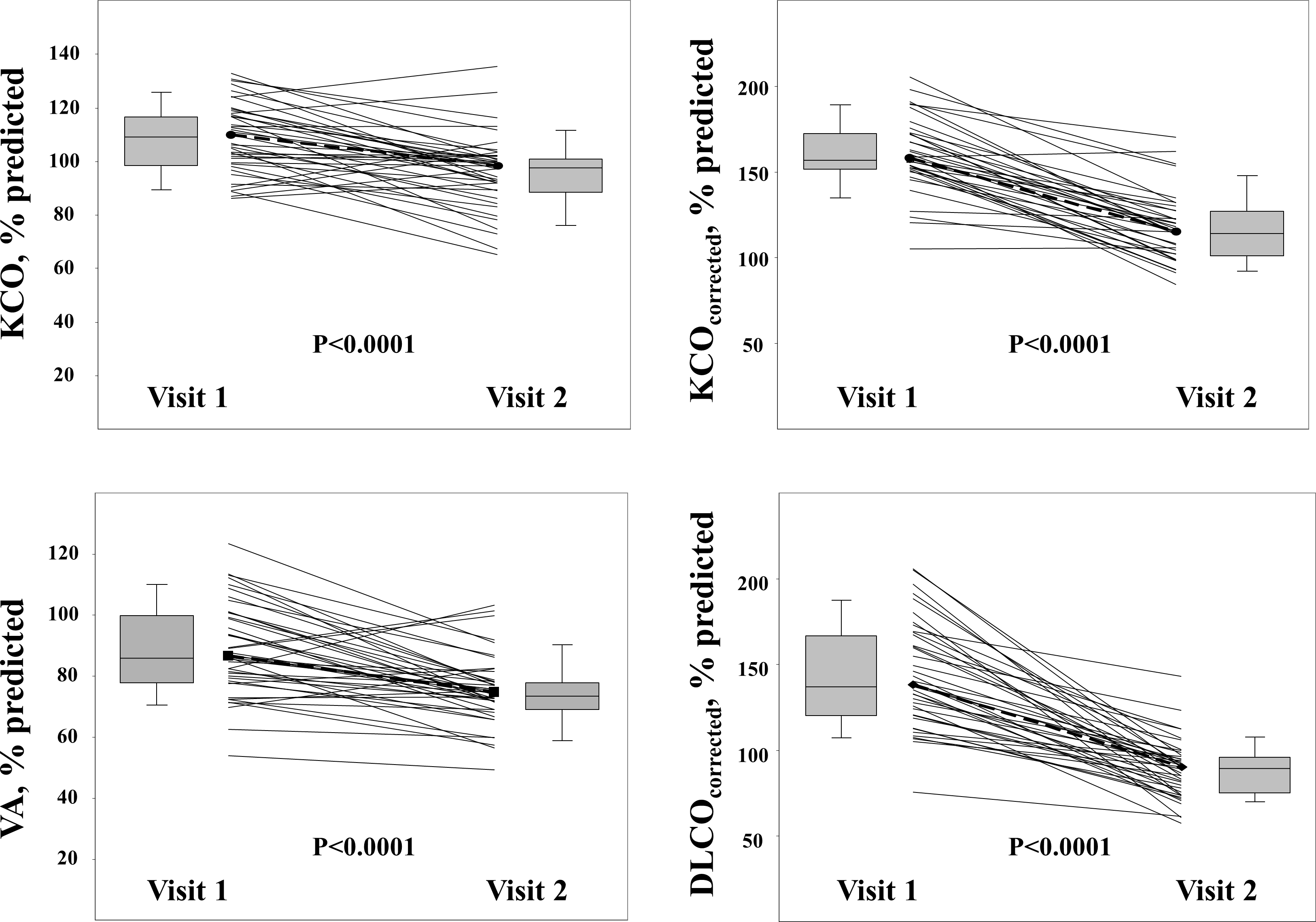
Deterioration of lung diffusion capacity during childhood in sickle cell diseaseWord count: 1500To the Editor,The American Society of Hematology guidelines, 2019, recommended obtaining pulmonary function tests (PFTs) in patients with sickle cell disease (SCD) with various respiratory symptoms even if they are at their steady state.1 These guidelines acknowledged that the usefulness of routine PFT is unknown because of the lack of research. However, this society further suggested that if the PFTs are obtained, it should be a comprehensive study including lung volumes and lung-diffusing capacity for carbon monoxyde (DLCO), in addition to spirometry.1 A large study in adult patients (n=310) with SCD showed that pulmonary function is abnormal in 90% of adult patients with Hb-SS.2 Common abnormalities included restrictive physiology and decreased DLCO. In this study, decreased DLCO indicated more severe sickle vasculopathy characterized by impaired hepatic and renal function, and a negative linear correlation existed between DLCO and age, suggesting that in adults with Hb-SS, disruption of alveolar–capillary gas exchange progressively deteriorated with time.2 Two recent cross-sectional studies of children with SCD showed that pulmonary function, including DLCO, worsened with age and showed correlations with biological markers of inflammation (induced sputum IL‐6 levels or blood neutrophilia).3,4Overall, these studies highlight the potential interest of DLCO measurement in SCD. The DLCO is the product of KCO (carbon monoxide coefficient of transfer) and alveolar volume (VA) and these two latter indices need to be interpreted separately since the decrease in DLCO is often mitigated by a preserved KCO or even increased KCO in SCD. It has been demonstrated that when corrected for hemoglobin levels, the children with SCD compared to controls of similar age had elevated KCOcorrected. The determination of alveolar-capillary membrane conductance (Dm) and pulmonary capillary blood volume (Vc) from the lung diffusing capacity for carbon monoxide (DLCO) or for nitric oxide (DLNO) has been done in SCD since the seventies, demonstrating an increase in Vc in SCD. KCO is mathematically linked to both Dm and Vc (1/KCO = VA/Dm + VA/θVc); thus, the increase in KCO is related to Vc increase, but since DLCO has been shown to worsen with age, the changes of DLCO, VA and KCO over time in children with SCD deserve to be studied.The objectives of our study were to describe the evolution of DLCO and its determinants, KCO and VA, and to further assess the initial risk factors of the decrease in DLCO in children/adolescents with SCD. To this end, we retrospectively recorded the routine follow-up PFTs of children with SCD who were included in a prospective cross-sectional study that included the measurement of both DLCO and DLNO with the calculation of Dm and Vc.5

Benjamin Dudoignon

and 8 more

Rationale: Patients with congenital central hypoventilation syndrome (CCHS) require long-term ventilation to ensure gas exchange and to prevent deleterious consequences for neurocognitive development. Two ventilation modes may be used for these patients depending on their tolerance, one invasive by tracheostomy and the other noninvasive (NIV). For patients who have undergone a tracheostomy, transition to NIV is possible when they meet predefined criteria. Identifying the conditions favorable for weaning from a tracheostomy it critical for the success of the process. Objective: The aim of the study was to share our experience of decannulation in a reference center; we hereby describe the modality of ventilation and its effect on nocturnal gas exchange before and after tracheostomy removal. Methods: Retrospective observational study at Robert Debré Hospital over the past 10 years. The modalities of decannulation and transcutaneous carbon dioxide recordings or polysomnographies before and after decannulation were collected. Results: Sixteen patients underwent decannulation following a specific procedure for transition from invasive to NIV. All decannulations were successful. The median age at decannulation was 12.6 [9.7; 15.0] years. Nocturnal gas exchange was not significantly different before and after decannulation, while expiratory positive airway pressure and inspiratory time increased significantly. An oronasal interface was chosen in two out of three patients. The mean duration of hospital stay for decannulation was 4.0 [3.0; 6.0] days. Conclusion: Our study underlines that decannulation and transition to NIV are achievable in CCHS children using a well-defined procedure. Patient preparation is crucial to the success of the process.

Plamen Bokov

and 4 more

Background. Whether small airway dysfunction (SAD), which is prevalent in asthma, helps to characterize wheezing phenotypes is undetermined. The objective was to assess whether SAD parameters obtained from impedance measurement and asthma probability are linked. Methods. One hundred and thirty-nine preschool children (mean age 4.7 years, 68% boys) suffering from recurrent wheeze underwent impulse oscillometry that allowed calculating peripheral resistance and compliance of the respiratory system (markers of SAD) using the extended RIC model (central and peripheral Resistance, Inertance and peripheral Compliance of the respiratory system). Children were classified using the probability-based approach of GINA guidelines (few, some, most having asthma). A principal component analysis (PCA) that determined the dimensions of wheezing disease evaluated the links between SAD and asthma probability. Results. Forty-seven children belonged to the few, 28 to the some and 64 to the most having asthma groups. Whereas their anthropometrics and measured parameters were similar, the most having asthma group exhibited the lowest mean value of airway inertance after bronchodilator probably due to airway inhomogeneities. PCA characterized nine independent dimensions including a peripheral resistance (constituted by baseline peripheral resistance, AX, R5-20Hz, X5Hz), a central resistance (baseline central resistance, R20Hz) and an airway size dimension (post-bronchodilator inertance and central resistance). PCA showed that the SAD markers were independent from clinical dimensions (control and asthma probability were two other dimensions) and did not help to define wheezing phenotypes. Conclusions. Lung function parameters obtained from impulse oscillometry and asthma probability were belonging to independent dimensions of the wheezing disease.

Bruno Mahut

and 3 more

Background: The Childhood Asthma Management Program study revealed that 25.7% of children with mild to moderate asthma exhibit a loss of lung function. The objective was to assess the trajectories of function by means of serial FEV1 in asthmatic children participating in out-of-hospital follow-up. Methods: A total of 295 children (199 boys) who had undergone at least 10 spirometry tests from the age of 8 were selected from a single-center open cohort. The annualized rate of change (slope) for prebronchodilator FEV1 (percent predicted) was estimated for each participant and three patterns were defined: significantly positive slope, significantly negative slope, and null slope (non-significant P-value in the Pearson test). The standard deviation (SD) of each individual slope was recorded as a variability criterion of FEV1. Results: The median (25th and 75th percentile) age at inclusion and the last visit was 8.5 (8.2, 9.3) and 15.4 (14.8, 16.0) years, respectively. Tracking of function (null slope) was observed in 68.8% of the children, while 27.8% showed a loss of function (negative slope) and 3.4% showed a gain in function (positive slope). The children characterized by loss of function depicted a better initial function and a lower FEV1 variability during their follow-up than children with tracking or gain of lung function. At the last visit, these children were characterized by a lower lung function than children with tracking or gain of lung function. Conclusion: Children with a better initial FEV1 value and less FEV1 variability are more prone to loss of lung function.