Gözde Nur Erkan

and 6 more

Objective: Dexmedetomidine has been shown to exert protective and curative effects on various tissues and organs in different pathological processes. This study aimed to investigate the effect of dexmedetomidine on the regeneration process after inducing a critical-sized bone defect in the calvarium of rabbits. Subject and Methods: Twenty-four male Oryctolagus cuniculus rabbits were divided into three groups, and an 8-mm circular parietal critical-sized bone defect was induced in all groups. Group_LD was given dexmedetomidine 2.75 µg/kg; Group_HD, dexmedetomidine 5.5 µg/kg; and Group_C, saline; all administered intraperitoneally for 7 days. The blood pressure and sedation score of the rabbits were evaluated. Bone tissue samples collected at the end of 8 weeks were examined via micro-computed tomography (micro-CT) and histomorphometry. Results: The micro-CT results indicated that regeneration significantly improved in all parameters in the dexmedetomidine-treated groups (p < 0.001). Furthermore, low-dose dexmedetomidine statistically significantly increased the bone volume ratio (BV/TV) compared with high-dose dexmedetomidine (p = 0.002). Trabecular thickness, connectivity value, and connectivity density were statistically significantly higher in Group_LD than in Group_HD (p < 0.001). The highest BA/TA% measurement in histomorphometry was observed in Group_HD, with a mean of 29.81% ± 8.52%. Significant intramembranous ossification was observed in the dexmedetomidine-treated groups, and active osteoblasts were observed in at the margin of the new bone trabeculae. Conclusion: This study demonstrated that dexmedetomidine increases osteoblastic activity and regeneration quality. In particular, low-dose dexmedetomidine exerted a more significant positive effect on the regeneration process and regenerative tissue quality than high-dose dexmedetomidine according to the micro-CT parameters.

Melek Volkan-Yazici

and 5 more

Background: Bruxism is significantly associated with craniofacial pain, feeling of stiffness or fatigue of the jaw and neck pain. Various physiotherapeutic strategies are used in the treatment of bruxism, however, it is not clear which method leads to greater decrease in pain. Objective: The aim of this study is to compare the effects of two physiotherapy methods (manual therapy (MT) and Kinesio taping with manual therapy (KTMT)) in patients with bruxism. Methods: Patients were randomized into MT or KTMT groups. Evaluations were performed at baseline and following four weeks of physiotherapy. Muscle thickness and stiffness were assessed via shearwave ultrasonography; pain thresholds were evaluated using algometer. Sleep quality was assessed using Pittsburgh Sleep Quality Index and Quality of life was assessed with Likert scales regarding the associated symptoms. Results: Significant improvements were found in muscle stiffness, pain threshold, sleep quality, quality of life (p<0.05) in both MT and KTMT group. Pain in bilateral temporalis and right occipital region of the trapezius muscle decreased more in the KTMT group compared to the MT group (p<0.05). No significant differences in muscle thickness (p>0.05) were found in either of the groups. Conclusion: Both MT and KTMT methods were effective in the treatment of bruxism. KT used in conjunction with MT has additionally decreased jaw pain and temporal region pain when compared to MT intervention only. Therefore, if jaw pain is the primary complaint of a patient, our results recommend including KT application in the physiotherapeutic treatment program.

Poyzan BOZKURT

and 2 more

Melike Başaran

and 6 more

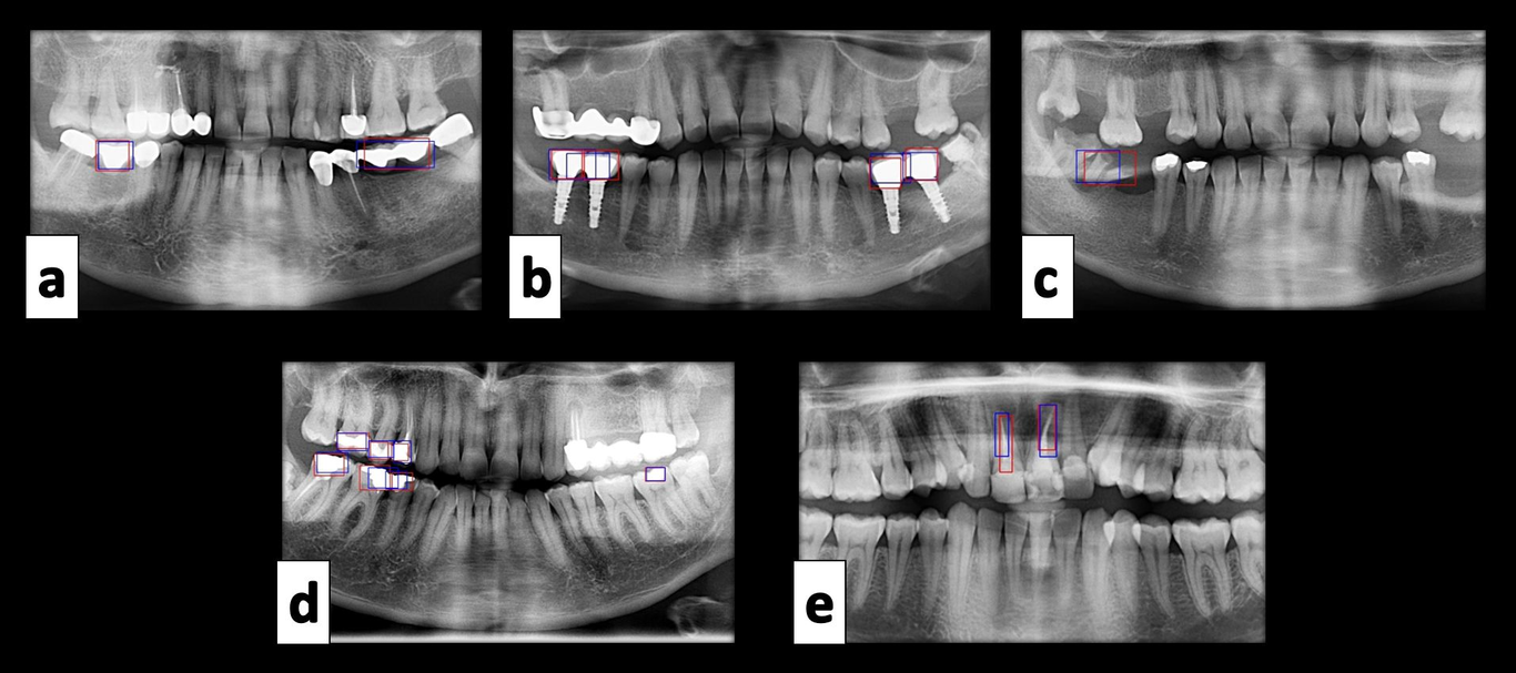
Aims of the Study: A radiographic examination is a significant part of the clinical routine for the diagnosis, management, and follow-up of the disease. Artificial intelligence in dentistry shows that the deep learning technique high enough quality and effective to diagnose and interpret the images in the dental practice. For this purpose, it is aimed to evaluate diagnostic charting on panoramic radiography using a deep-learning AI system in this study. Methods: 1084 anonymized dental panoramic radiographs were labeled for 10 different dental situations including crown, pontic, root-canal treated tooth, implant, implant-supported crown, impacted tooth, residual root, filling, caries, and dental calculus. AI Model (Craniocatch, Eskişehir, Turkey) based on a deep CNN method was proposed. A Faster R-CNN Inception v2 (COCO) model implemented with Tensorflow library was used for model development. The training and validation data sets were used to predict and generate optimal CNN algorithm weight factors. Results: The proposed artificial intelligence model has promising results for detecting dental conditions in panoramic radiographs except for caries and dental calculus. The most successful F1 Scores were obtained from the implant, crown, and implant-supported crown as 0,9433, 0,9122, 0,8947, respectively. Conclusion: Thanks to the improvement of the success rate of AI models in all areas of dentistry radiology, it is predicted that they will help physicians especially in panoramic diagnosis and treatment planning, as well as digital-based student education, especially in this pandemic period when online training is on our agenda.