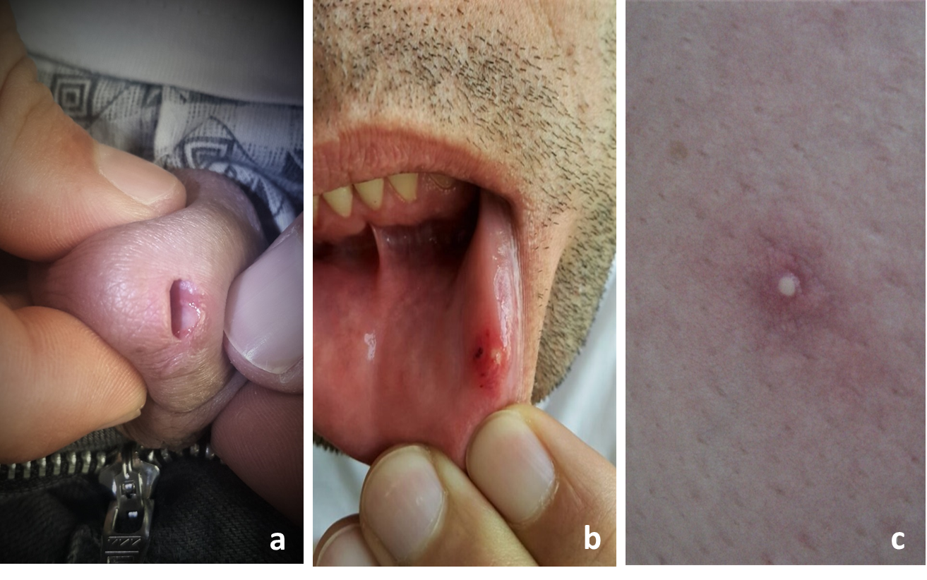
An unusual localization of genital ulcer in Behçet’s disease: external urethral meatusRunning head: Unusual genital ulcer in Behçet’s diseaseÇağrı Turan1, İbrahim Karabulut 21Department of Dermatology and Venereology, the Republic of Turkey, Health Sciences University Erzurum Regional Training and Research Hospital, Turkey2Department of Urology, the Republic of Turkey, Health Sciences University Erzurum Regional Training and Research Hospital, TurkeyCorresponding Author : Çağrı TURAN; Department of Dermatology and Venereology, the Republic of Turkey, Health Sciences University Erzurum Regional Training and Research Hospital, Turkey; Üniversite Mahallesi, Çat Yolu Cd., Yakutiye/Erzurum, Post Code: 25070e-mail: [email protected], telephone number: +905445252504Word count: 471Table count: 0Supplementary table: 0Figure count: 1Funding sources : We declare no financial support or relationships that may pose a conflict of interest.Conflict of interest: There is no conflict of interest.The paper has not been published or submitted for publication elsewhere.All authors have contributed significantly, and all authors agree with the content of the manuscript.Informed consent form was obtained from the patient.Key words: Behçet’s disease, genital ulcer, Urethra, VasculitisDear editor,Behçet’s disease (BD) is a chronic, recurrent, multisystemic vasculitis which can affect all vascular system. The most common symptom is genital ulcer accompanying oral aphthae, and its diagnosis is currently made according to the International Criteria for Behçet’s Disease (ICBD). Providing two points for oral aphthae, genital ulcers, and ocular involvement and one point for the other skin lesions (erythema nodosum, papulopustular/acneiform lesions in post-adolescent), vascular involvement, and neurological findings are evaluated if the patient scores reach four or more; the patient is considered to be BD1, 2. In men, genital aphthous ulcers occur in 60 to 65% of cases and are most common in the scrotum, shaft and glans penis, and rarely in the groin and perineum, extremely rare in the urethral orifice2, 3We presented a patient with a complaint of painful micturition, ultimately diagnosed with BD. A 34-year-old male patient was consulted from the urology following the evaluation of his painful voiding complaint 10 days accompanying wound in the periurethral orifice. On genital examination, an oval, and sharp circumscribed aphthous ulcer with a serous floor, approximately 4 mm in diameter, was seen on the external urethral orifice (Figure 1a). The patient who had no known disease has refused to use any medication, similar complaints, suspicious sexual contact, except for oral aphthae recurring 8-10 times a year (Figure 1b). After noticing a few pustular lesions with peripheral partial erythema on his back, we focused on BD and inflammatory bowel diseases in the examination (Figure 1c). We learned that the patient had no family history and other related symptoms. Bowel habits were normal. Hepatitis, HIV, syphilis serologies were negative. CRP and sedimentation were 3.2 mg/dl (0-5 mg/dl) and 27 mm/hour (0-20 mm/hour); respectively. Other hematological and biochemical parameters were within normal limits. Complement levels, antinuclear antibody, anti-dsDNA, p-ANCA, c-ANCA and rheumatoid factor were normal. Pathergy test was positive. The patient was diagnosed with BD with a score of 6 according to the current ICBD, and no pathology was found in the eye and cardiology consultations. His complaints improved within 1 week without leaving any scar, after treatment with topical betamethasone valerate applied to the urethral orifice and oral colchicine 1.5 mg/day. No clinical progression has been observed for 2 years in the follow-up.A urethral ulcer is an unusual finding in BD. Aktaş recently reported a 27-year-old male patient with clinical features quite similar to our case, but with negative pathergy4. Interestingly, Cobilinschi et al. reported that a 34-year-old female patient with BD who was admitted with progressive dysuria, pain in the right lumbar region, and hydronephrosis was determined a necrotic ulcer in the ureter whose histopathology was compatible with vasculitis5.We present this case to draw attention to the aphthous ulcer in the urethra as an unusual genital involvement for BD, and the importance of physical examination.