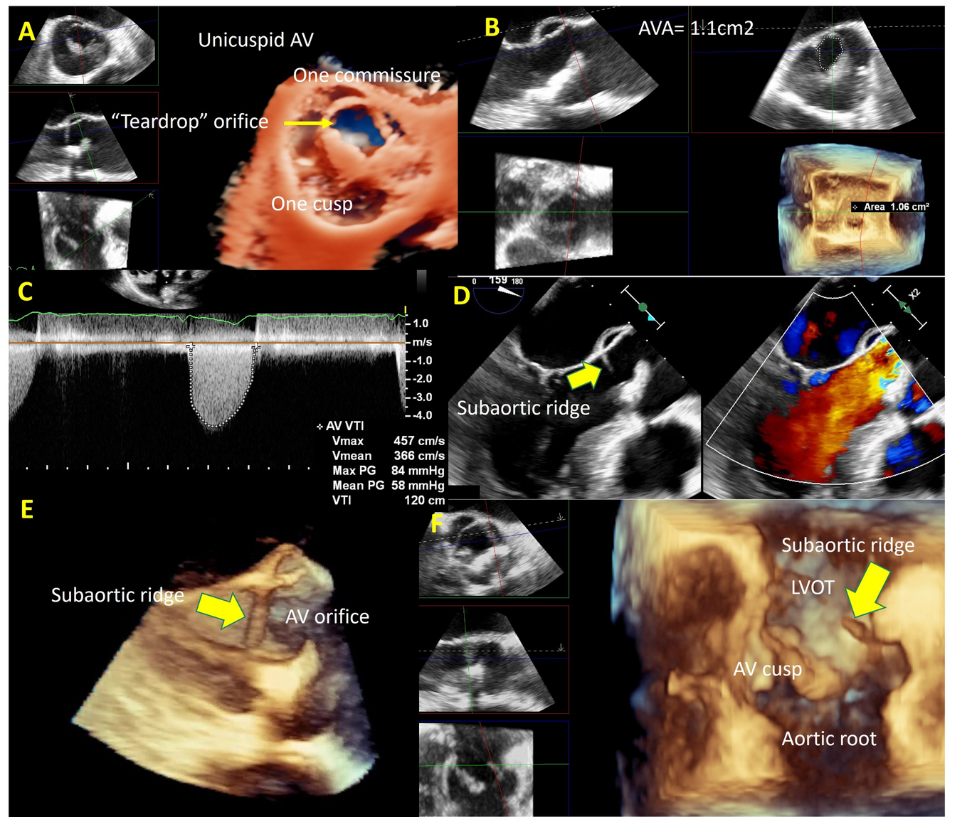
Aims To compare the effects of LV volume overload due to chronic organic MR or AR on RV shape and function. Methods and results We studied 63 patients with moderate-severe or severe primary MR and 36 patients with moderate or severe AR. 3D echocardiography of LV and RV was performed to measure volumes and EF. RV fractional area change (FAC) was calculated, and RV shape was assessed by calculating the RV eccentricity index.LV EDVi was significantly larger in the patients with AR than in those with MR. RV EF and RV FAC were lower in pts with AR than in those with MR. RV EI was significantly higher in the AR group. In both groups, LV EDVi showed positive correlations with RV EI ( r= 0.693 for AR and r=0.399 for MR) and negative correlations (RV EF :r= -0.545 for AR and r=- 0.383 for MR ; RV FAC: r=-0.816 for AR and r=-0.647 for MR, ). LV sphericity index showed negative correlations (RVFAC: r= -0.512 and r=-0.608 f ;RV EF:r=-0.408 and r=-0.469 respectively ) and positive correlation with the RV EI (r= 0.39 and r=0.511 respectively) .LV EDVi and LV sphericity index were found to be the only independent predictors of RV eccentricity index, EF, and FAC. Conclusions RV remodeling in chronic LV overload due to MR or AR occurs independently on PASP values. LV size and shape are the only independent predictors of RV geometry and function. Accordingly, chronic AR has a greater impact on RV than MR