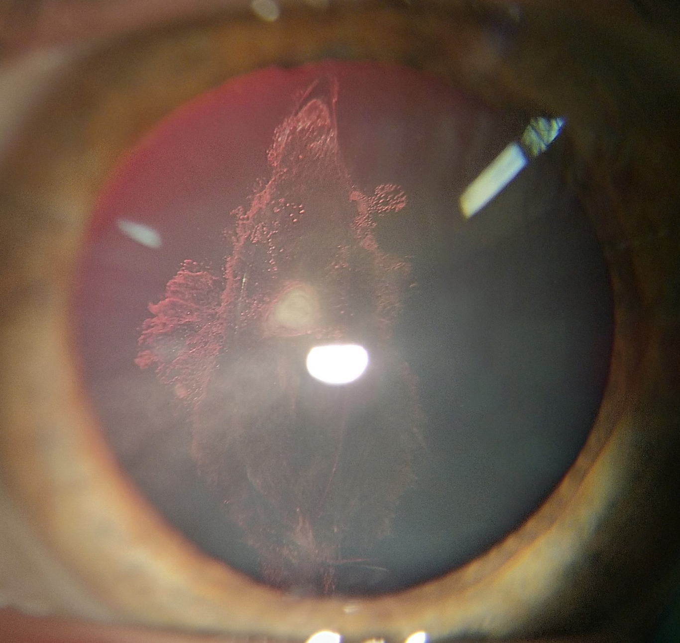
RECALCITRANT MULTIDRUG-RESISTANT PSEUDOMONAS KERATITIS WITH SUBSEQUENT TRIPLE PROCEDUREAuthors: Ēriks Elksnis1,2,4, Eva Elksne1,3, Olita Lūse1,2, Juris Vanags1,4, Guna Laganovska1,4Affiliations:Riga Stradins University, Riga, LatviaLatvian American Eye CenterChildren’s Clinical University Hospital, Riga, LatviaPauls Stradins Clinical University HospitalCorrespondence to: Ēriks Elksnis. ORCID 0000-0002-7899-8224Riga Stradins University, Dzirciema street 16, Riga, Latvia. LV1007.Data availability statement : We hereby transfer, assign, or otherwise convey all copyright ownership, including any and all rights incidental thereto, exclusively to the journal, in the event that such work is published by the journalConflict of interest disclosure : Authors declare no potential conflicts of interest.Ethics approval statement : All procedures performed involving this case were in accordance with the ethical standards of the institutional research committee and with the 1964 Helsinki declaration and its later amendments or comparable ethical standards.Consent statement: Written informed consent was obtained from the patient to publish this report in accordance with the journal’s patient consent policy.KEY CLINICAL MESSAGERecalcitrant Pseudomona aeruginosa keratitis is a challenging case in ophthalmology and can lead to irreversible blindness if not treated properly and in time.KEYWORDS: Pseudomona aeruginosa, recalcitrant keratitis, penetrating keratoplasty.INTRODUCTIONCorneal opacity is the 5th leading cause of blindness and visual impairment, affecting approximately 6 million people worldwide; additionally, it is responsible for 1.5–2.0 million new cases of monocular blindness per year. Among all etiologies (such as infection, trauma, and inflammation), infectious keratitis (IK) is the main cause of corneal blindness, with an estimated incidence ranging from 2.5–799 per 100,000 population years (Ting et al. 2021).Pseudomonas aeruginosa (P. aeruginosa ) is a gram-negative aerobic pathogen that can cause a wide range of infections and is one of the main causative pathogens of bacterial keratitis, especially in contact lens-associated keratitis, potentially leading to sight-threatening complications if not appropriately treated (Hilliam, Kaye & Winstanley 2020).A particular characteristic of multidrug resistant Pseudomonas keratitis is its rapid progression, in which corneal destruction can be completed within 24–48 hours in some of the more virulent bacterial strains (Reynolds & Kollef 2021). Therefore, P. aeruginosa -caused keratitis is often associated with a high-cost long treatment period and poor visual outcomes (Hilliam, Kaye & Winstanley 2020).CASE PRESENTATIONA 60-year-old female presented to a tertiary medical center in May 2021 with progressive vision loss, purulent discharge, photophobia, foreign body sensation, and pain in the left eye (LE).Three weeks prior, the patient developed a foreign body sensation in her LE due to contact lenses remaining unremoved for one night. She had visited her local ophthalmologist and been prescribed topical tobramycin/dexamethasone 3 mg/mL four times daily, cyclopentolate 10 mg/mL twice daily, and dexpenthenolum once daily before bedtime. There was clinical improvement during the first 5 days of therapy; however, severe clinical worsening followed. The patient returned to the outpatient department 8 days after the first consultation, and treatment was changed to topical chloramphenicol/dexamethasone 1 mg/2 mg/mL six times daily, cyclopentolate 10 mg/mL twice daily and oral doxycycline 100 mg twice daily. Despite treatment, the symptoms worsened in the following week.On the presenting day at the tertiary medical center, the best corrected visual acuity (BCVA) was 20/25 in the right eye (RE) and hand-motion (HM) vision in the LE. The RE was unremarkable on initial examination, whereas slit-lamp examination of the LE revealed a massive amount of purulent discharge in the conjunctival fornixes and intense mixed conjunctival injection. A large epithelial defect in the cornea was present, associated with a ring-like stromal infiltrate 3.1 mm in height, which was “soupy” in appearance owing to stromal necrosis. A hypopyon 3.2 mm in height was observed (Figure 1A). Both the lids were swollen and erythematous. As the posterior segment could not be visualized, B-scan ultrasonography of the LE was performed and confirmed a flat retina with no vitritis.Corneal scrapings and cultures were obtained from LE. Cultures were acquired using a sterile cotton-tipped swab and placed in transport medium. The scrapings were placed on a glass slide and, together with the culture, forwarded to a microbiology laboratory.Topical chloramphenicol/dexamethasone was discontinued, and levofloxacin 5 mg/mL eyedrops were initially administered every hour. Additionally, cyclopentolate 10 mg/mL was administered twice daily to control pain and prevent synechia, artificial tear drops were administered to aid wound healing, and oral doxycycline 100 mg twice daily was administered to prevent keratolysis.After 12 hours, the condition worsened; discharge remained in the same amount, however, the hypopyon increased to 5.4 mm in height, with an infiltration spread 360 °around the limbus and significant corneal edema (Figure 1B).Levofloxacin was switched to moxifloxacin 5 mg/mL eye drops every hour. As a result, the amount of purulent discharge reduced, yet the central corneal infiltrate and ulcer remained unchanged, with remarkable stromal necrosis and corneal thinning in the nasal limbs (Figure 1C).On the fourth day from admission, multidrug resistantP.aeruginosa was isolated from the culture. (Table 1). Based on these results, and after consultation with an infectologist, moxifloxacin was changed to fortified piperacillin 1.5 mg/mL/tazobactam 12 mg/mL eye drops every hour.