Eric Jauniaux

and 2 more

Aortic balloon for the intraoperative management of placenta accreta spectrum: need for standardised methodology and safety dataIn 2003, Bell-Thomas et al (BJOG 2003;110:1120-1122) reported on the emergency use of a transfemoral aortic occlusion catheter to control massive haemorrhage in a case of caesarean hysterectomy for placenta percreta. This was only the second case published in the international literature on the use of an intra-abdominal aortic balloon occlusion (IABO) in the management of placenta accreta spectrum (PAS) (Paull et al. Anesth Intensive Care 1995;23:731-734). A non-exhaustive Pub-Med literature review of articles published in English on this the topic over the last 20 years, identifies 27 articles, 23 of which come from the Peoples Republic of China, where IABO seems to be increasingly popular in the management of PAS.In brief, IABO involves the insertion of a balloon catheter into the infrarenal abdominal aorta above the aortic bifurcation under fluoroscopy guidance. The procedure is performed in a hybrid operating room or interventional radiology (IR) suite with secondary transfer to the operating room. In all but one study (Zhu et al Biomed Res Int.2017:8604849), the balloon was inflated after delivery of the newborn.All publications so far have been retrospective and most are case-control studies, comparing the outcomes of IABO with those of routine surgical techniques with or without additional procedures such as intra-uterine tamponade. Recently, authors have also started to compare IABO with iliac artery balloon occlusion.Overall, these studies have shown that IABO is associated with reduced estimated blood loss and transfusion requirement, ICU admission and hysterectomy and suggested that IABO is more effective than iliac artery balloon occlusion, presumably as arterial occlusion is more effective.However, there is wide variation between studies in prenatal imaging and clinical selection criteria, intraoperative IR methodology and confirmation of the diagnosis of PAS at birth. For example, the pre-operative fluoroscopy time ranges between 2 and 25 minutes with fetal radiation exposure of 4 to 25 mGy; intraoperative balloon inflation/deflation time varies between 5-10/1 minutes and 45-80/10 minutes. The size of the balloon and the need for transfer between the IR room and the operative theatre is rarely described. Most studies lack histopathology confirmation of the diagnosis and/or stratification by PAS grade.Heterogeneity in methodology and design leads to a high risk of confounding, bias or chance. There is also a high risk that the relationship is not causal. One major concern is the risks-benefit ratio of the use of IABO for both mothers and fetuses, in particular if they do not have PAS. The most commonly reported post-operative complication associated with IABO are arterial thrombosis of the external iliac or the femoral artery. There are no data on the long-term follow of the children born after IABO.In 2018, the expert panel of the RCOG green top guidelines 27a (Jauniaux et al., BJOG.2019;126:e1-e48) concluded that larger studies are necessary to determine the safety and efficacy of IR before this technique can be advised in the routine management of PAS. The 10 new studies published in 2019-2010 on the use of IABO in the management of PAS are insufficient to change this statement.Word count: 499

Eric Jauniaux

and 1 more

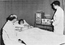
The prenatal diagnosis of fetal anomalies started with the development of X-ray. In 1943, Hartley and Burnet, Radiologists in Manchester (J Obstet Gynaecol Brit Empire,1943;50:1-12), reported a series of 11 cases of “croaniolacunia” or lacunar skull, a condition often associated at births with spina bifida or encephalocele. These cases were all diagnosed in the third trimester of pregnancy and the radiograph features believed to be due to the effect of increased intracranial pressure on the fetal skull of hydrocephaly. Until the end of the 1960s, radiography remained the main technique to diagnose congenital abnormalities. In 1969, Russell (J Obstet Gynaecol Br Commonw,1969;76:345-50), also a consultant radiologist from Manchester, compared the accuracy of antenatal radiology examinations with paediatric reports in the diagnosis of anencephaly and other major neural tube defects, skeletal abnormalities such as achondroplasia and severe exomphalos when associated with rib deformities. Overall, the accuracy of the radiological diagnosis was considered as “strikingly” accurate for neural tube defects with 88 out of 113 cases of anencephaly diagnosed before delivery. Although, the author did not provide the gestational age at diagnosis, the images included in the article indicate that these were obtained in the third trimester. As neonatal care and surgery were in their infancy at the time, the main objective in diagnosing these anomalies was not the fetus but the need to identify antenatally mothers at risk of obstructed labour.Not surprisingly, some of the first publications by the team of Ian Donald in Glasgow were on the antenatal use of ultrasound imaging in the evaluation of the size of the fetal head (Willocks et al., J Obstet Gynaecol Br Commonw,1964;71:11-20). The fetal head was the only structure that could be measured and biparietal diameter the only measurement that could be obtained with the “ultrasound beam” of the first ultrasound machine (Figure). The technique called “cephalometry” was used at the end of the third trimester to assess “growth and maturity” of the fetus and “disproportion” and was found to be more reliable with ultrasound than X-ray. It would be another decade, before ultrasound imaging could reliably identify fetal anomalies such as spina-bifida in the second trimester of pregnancy (Campbell et al., Lancet,1975;1(7920):1336-7). However, the use of ultrasound imaging in the mid-seventies to search for major neural tube defects was always triggered by high levels of maternal serum alpha-fetoprotein. As there were few ultrasound equipment available and few trained operators, this biomarker was to remain for two decades the first line of action in the antenatal screening strategy for spina-bifida. The advent of high-resolution imaging, access low-cost and mobile ultrasound equipment and the training of more specialists and sonographers has moved the antenatal screening and diagnosis of many fetal anomalies to 11-14 weeks of gestation (Ushakov et al., UOG,2019;54:740-5).The systematic review by Drukker et al. (BJOG 2020) brings the focus back to late pregnancy: even in our exciting modern era of early anomaly scanning, a fetal abnormality will still be found in about 1 in 300 women scanned in the third trimester.Word count: 500BJOG since 1902 Perspectives on BJOG-20-0525R1