BACKGROUND: Increased systolic pulmonary artery pressure (sPAP) could lead to mechanical dysfunction and myocardial fibrosis of right heart chambers. Echocardiographic strain analysis has not been adequately studied in patients living with pulmonary hypertension (PH). METHODS AND RESULTS: A cross-sectional cohort of patients with suspected PH and echocardiographic strain evaluation was recruited. Cut-off values of peak tricuspid regurgitation velocity with low probability of PH (≤ 2.8 m/s), intermediate probability (2.9-3.4 m/s, without other echo PH signs) and high probability of PH (2.9-3.4 m/s with other echo PH signs and >3.4 m/s) categories were studied by right ventricular and right atrial strain analysis in a sample of 236 patients, 58 (56.9%) had low, 15 (14.7%) intermediate, and 29 (28.4%) high probability of PH. We observed a negative association between right ventricular free wall strain and atrial global strain with sPAP. As PH severity increased, right atrial reservoir, conduit, and contraction (booster) strain values decreased. Identified cut-off values of strain parameters had an adequate ability to detect PH severity categories In addition, post-mortem biopsies of right heart chambers from subjects with known severe pulmonary hypertension were analyzed to quantify myocardial fibrosis. Our sample of right heart biopsies (n=12) demonstrated an association between increased sPAP before death and right ventricular and right atrial fibrosis. CONCLUSIONS: Mechanical dysfunction and fibrosis in right chambers is associated with increased sPAP. Right ventricular and atrial strain could provide enhancement in the diagnosis and categorization of subjects with suspected PH.

R.B SINGH

and 12 more

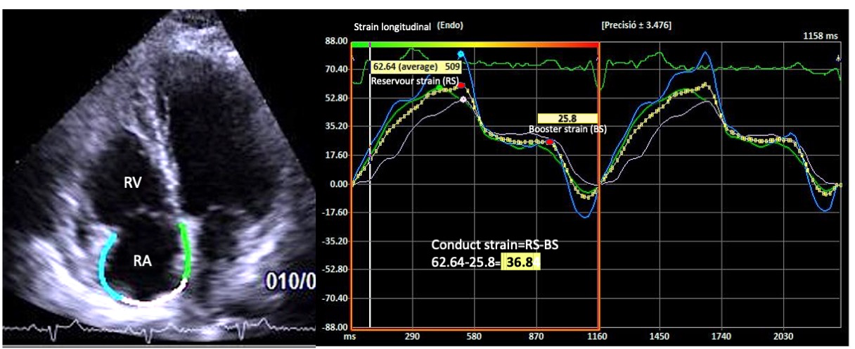
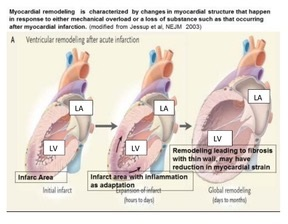
Chronic heart failure (CHF) has different stages and they include pre-HF (PHF), a state of high risk of developing myocardial dysfunction and subsequently, advanced CHF (ACHF). Some major behavioral risk factors of PHF are Western diet, tobacco, alcoholism, sedentary life style and chronic mental disorders (anxiety, depression). These insults might predispose to biological risk factors such as obesity, diabetes mellitus dyslipidemia hypertension, myocardial infarction and cardiomyopathy. These risk factors damage the myocytes leading to fibrosis, apoptosis, cardiac hypertrophy, along with alterations in cardiomyocyte’ size and shape. A condition of physiological subcellular remodeling resulting into a pathological or deformation state might be developed conductiong to PHF. Both PHF and HF are associated with the activation of phospholipases and protease, mitochondrial dysfunction, oxidative stress and development of intracellular free Ca2+ [Ca2+]i overloading or an elevation in diastolic [Ca2+]i. Simultaneously, cardiac gene expression is activated leading to further molecular, structural and biochemical changes of the myocardium. The sub-cellular remodeling may be intimately involved in the transition of cardiac hypertrophy to heart failure. Two-dimensional (2D) and three-dimensional (3D) speckle tracking-echocardiography (STE) have been used to quantify regional alterations of longitudinal strain and area strain, through their polar projection, which permits a further assessment of both sites and degrees of myocardial damage. The examination of strain can identify sub-clinical cardiac dysfunction or cardiomyocyte remodeling. It is concluded that during remodeling of the myocardium, cardiac strain is attenuated which can be used for the assessment of disease progression and subsequently for therapeutic interventions.

Kirolos Barssoum

and 12 more

Background: Right ventricular failure (RVF) following Left Ventricular Assist Device (LVAD) implantation is associated with worse outcomes. Prediction and early identification of RVF with speckle-tracking echocardiography (STE) has been proposed. Methods: We queried multiple databases for articles reporting on pre-operative/intraoperative global longitudinal strain (GLS) and free-wall strain (FWS) in LVAD recipients. We performed a systematic review and meta-analysis of published literature. The standard mean difference (SMD) in GLS and FWS in patients with and without RVF postoperatively was pooled using random effects model. Results: Fifteen studies, with a total of 967 LVAD recipients were included. There was statistically significant difference in GLS among patients who did and did not develop RVF; SMD= -3.09 (95% CI: -4.62 to -1.57; p-value <0.0001). There was significant difference in FWS between two groups; SMD: -2.75 (95% CI: -3.72 to -1.79; p-value <0.0001). Upon subgroup analysis of imaging modality, transthoracic echocardiography (TTE)-derived GLS and FWS remained predictive for RVF with SMD of -3.97 (95% CI: -5.40 to -2.54; p-value <0.001) and -3.05 (95% CI: -4.11 to -1.99; p-value <0.001), respectively. However, there was no significant difference between RVF and non-RVF groups upon using transesophageal echocardiography (TEE) to assess GLS and FWS. Conclusion: GLS and FWS assessment of the RV by STE is a useful tool to predict postoperative RVF in LVAD recipients. While the predictive role of TTE was robust, the TEE-derived measures seemed to be less predictive. Future studies need to specify the strain cut-off value that can predict the adverse outcome of RVF

Kirolos Barssoum

and 15 more

Background This meta-analysis aims to evaluate the utility of speckle tracking echocardiography (STE) as a tool to evaluate for cardiac sarcoidosis (CS) early in its course. Electrocardiography and echocardiography have limited sensitivity in this role, while advanced imaging modalities such as cardiac magnetic resonance (CMR) and 18F-Fluorodeoxyglucose–Positron Emission Tomography (FDG-PET) are limited by cost and availability. Methods We compiled English language articles that reported left ventricular global longitudinal strain (LVGLS) or global circumferential strain (GCS) in patients with confirmed extra-cardiac sarcoidosis versus healthy controls. Studies that exclusively included patients with probable or definite CS were excluded. Continuous data were pooled as a standard mean difference (SMD) between the sarcoidosis group and controls. A random effect model was adopted in all analyses. Heterogeneity was assessed using Q and I2 statistics. Results Nine studies with 967 patients were included in our analysis. LVGLS was significantly lower in the extra-cardiac sarcoidosis group as compared to controls, SMD -3.98, 95% confidence interval (CI): -5.32, -2.64, p< 0.001, also was significantly lower in patients who suffered Major Cardiac Events(MCE), -3.89, 95% CI -6.14, -1.64, p< 0.001 . GCS was significantly lower in the extra-cardiac sarcoidosis group as compared to controls, SMD: -3.33, 95% CI -4.71, -1.95, p< 0.001 Conclusion LVGLS and GCS were significantly lower in extra-cardiac sarcoidosis patients despite not exhibiting any cardiac symptoms. LVGLS correlates with MCEs in CS. Further studies are required to investigate the role of STE in the early screening of CS.