Craig Basman

and 10 more

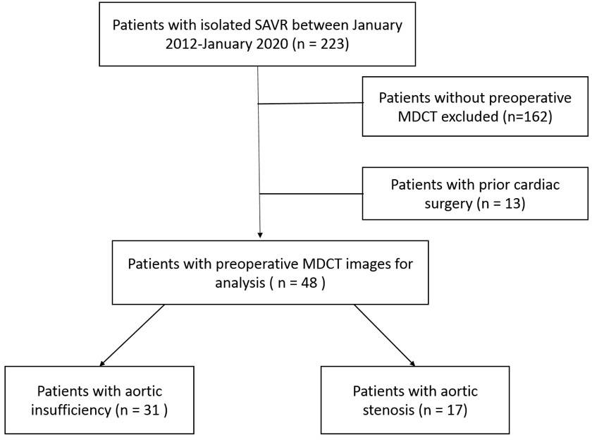
Background: Transesophageal echocardiogram (TEE) is the gold-standard for diagnosis of degenerative mitral regurgitation (MR) and is used for guidance of transcatheter mitral valve repair (TMVr). However, TEE is an invasive diagnostic modality that requires anesthesia and esophageal intubation. Multi-detector computed tomographic angiography (MDCT) provides high resolution images and three dimensional (3D) reconstructions that allow for comprehensive assessment of complex mitral anatomy. We hypothesized that MDCT can yield similar information to 3D TEE relevant to TMVr, deferring the need for a preoperative TEE. Methods: Patients that underwent TMVr (or were evaluated for transcatheter mitral valve replacement) for degenerative MR were retrospectively analyzed from 2017 to 2019 at a single center. Patients were included in the analysis if preoperative MDCT was performed. Two experienced TEE and two MDCT readers, blinded to patient outcome and alternative imaging modality, analyzed the following characteristics: leaflet pathology (flail, degenerative, mixed), leaflet location (A1-3/P1-3), mitral valve area (MVA), flail width/gap, anterior-posterior (AP) and commissural diameters, posterior leaflet length, leaflet thickness, presence of mitral valve cleft and degree of mitral annular calcification (MAC). Results: Of the 87 patients, 22 had preoperative MDCT. MDCT was able to correctly identify the leaflet pathology in 77% (17/22). Eleven patients had a flail leaflet with 91% (10/11) identified on MDCT and MDCT correctly predicted the dysfunctional leaflet location in 95% (21/22). Measurements were not significantly different for MVA, flail width, commissural diameter, AP diameter, posterior leaflet length and leaflet thickness. However, measurements on MDCT were significantly overestimated for flail gap compared to TEE. Degree of MAC was similar in 91% (10/11) between imaging modalities. Conclusion: MDCT provides similar measurements to 3D TEE for preoperative TMVr planning. Further studies are required to establish novel imaging algorithms utilizing MDCT to reduce the need for preoperative TEE.

Craig Basman

and 8 more

ABSTRACT: Background: Valve-in-valve (ViV) transcatheter aortic valve replacement (TAVR) has become a valuable option in patients with bioprosthetic failure. However, potential issues with ViV TAVR may occur in patients with high risk anatomy for coronary obstruction and patients with baseline smaller bioprosthetic valves at risk for patient prosthesis mismatch. The purpose of this study was therefore to use preoperative electrocardiography (ECG)-gated, multidetector computed tomography (MDCT) in patients undergoing isolated surgical aortic valve replacement (SAVR) to 1) identify which would be high risk for coronary occlusion with ViV TAVR, and 2) predict intraoperative SAVR sizing. Methods: Among 223 patients from our institutions’ database that underwent SAVR for aortic insufficiency (AI) or aortic stenosis (AS) between January 2012 and January 2020, 48 patients had MDCT imaging prior to surgery (AI; n=31, AS; n=17). Of all patients, 67% (n=32) were bicuspid morphology. Results: With the use of virtual valve implantation, all patients with AI and bicuspid AS had feasible anatomy for ViV TAVR, while 38% of patients with tricuspid AS were high risk for coronary obstruction. There was a strong correlation between actual valve size implanted and preoperative MDCT measurements using annulus average diameter, area and/or perimeter. Conclusion: Preoperative MDCT in patients undergoing SAVR is a useful tool for lifetime management, particularly in patients with tricuspid AS. Decisions for surgical management may change based on MDCT’s ability to predict intraoperative SAVR size and determine which patients may be high risk candidates for future ViV TAVR due to coronary artery obstruction.

Craig Basman

and 9 more

Background: Hybrid coronary revascularization (HCR) constitutes a left internal mammary artery (LIMA) graft to the left anterior descending (LAD) coronary artery, coupled with percutaneous coronary intervention (PCI) for non-LAD lesions. This management strategy is not commonly offered to patients with complex multi-vessel disease. Our objective was to evaluate 8-year survival in patients with triple-vessel disease (TVD) treated by HCR, compared with that of concurrent matched patients managed by traditional coronary artery bypass grafting (CABG) or multi-vessel PCI. Methods: A retrospective review was undertaken of 4805 patients with TVD who presented between January 2009 and December 2016. A cohort of 100 patients who underwent HCR were propensity-matched with patients treated by CABG or multi-vessel PCI. The primary end-point was all-cause mortality at 8 years. Results: Patients with TVD who underwent HCR had similar 8-year mortality (5.0%) as did those with CABG (4.0%) or multi-vessel PCI (9.0%). A composite end-point of death, repeat revascularization, and new myocardial infarction, was not significantly different between patient groups (HCR 21.0% vs. CABG 15.0%, p = 0.36; HCR 21.0% vs. PCI 25.0%, p = 0.60). Despite a higher baseline SYNTAX score, HCR was able to achieve a lower residual SYNTAX score than multi-vessel PCI (p = 0.001). Conclusions: In select patients with TVD, long-term survival and freedom from major adverse cardiovascular events (MACE) after HCR are similar to that seen after traditional CABG or multi-vessel PCI. HCR should be considered for patients with multi-vessel disease, presuming a low residual SYNTAX score can be achieved.